
Parvoviridae
Read
Table of Contents
Viral Characteristics
Classification
Parvovirus
Feline Panleucopenia
Canine Parvovirus Infection
Porcine Parvovirus Infection
Mink and Raccoon Enteritis
Goose Parvovirus Infection
Canine Minute Virus
"AMDV-like viruses"
Aleutian Mink Disease
"BPV-like viruses"
Bovine Parvovirus Infection
Glossary
This family consists of small non-enveloped DNA viruses that infect both vertebrates and arthropods. It includes a number of important veterinary pathogens.
Viral Characteristics
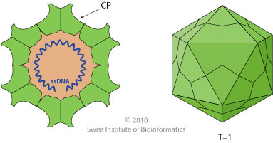
Very small (20 - 22 nm), non-enveloped, single-stranded DNA viruses with icosahedral symmetry (See Fig. 9.1).
The virions are replicated in the nuclei of rapidly dividing cells.
The ssDNA serves as a template for synthesis of dsDNA by host enzymes. The dsDNA then serves as a template for the production of mRNA and progeny genomes.
The progeny viruses are assembled in the nucleus; host cell replication results in cell lysis.
The human B19 parvovirus (erythema infectiosum, aplastic anemia) replicates only when a cell is in S phase of the cell cycle. This accounts for why it replicates in red blood cell precursors rather than in mature red blood cells.
Virions are stable in the environment, resisting heat, desiccation and some disinfectants. Aqueous sodium hypochlorite solution is effective against this and other viruses.
Some species of parvoviruses agglutinate red cells (hemagglutination) a characteristic utilized in some diagnostic procedures. These hemagglutinating viruses have an erythrocyte attachment protein (hemagglutinin) on their surface.
The pathogenesis of the parvovirus infections described below is in general similar; see feline panleukopenia.
Figure 9-1. Illustration of Parvoviridae (20 - 22 nm) capsid.
Classification
The family Parvoviridae has two subfamilies, Parvovirinae and Densovirinae.
Subfamily Parvovirinae has three genera and two generic groups. The important veterinary viruses in these categories are as follows:
Parvovirus:
Feline panleucopenia virus
Canine parvovirus
Porcine parvovirus
Virus of mink and raccoon enteritis
Goose parvovirus
Canine minute virus
Erythrovirus
B19 - Human parvovirus (causes erythema infectiosum in children)
Dependovirus:
Consists of defective viruses that require an adenovirus (or herpesvirus) "helper" in order to replicate efficiently. They are also called AAV (adeno-associated viruses). They are clinically unimportant, yet are being considered as vectors for gene therapy.
"AMDV-like viruses"
Aleutian mink disease virus
"BPV-like viruses"
Bovine parvovirus
Subfamily Densovirinae (three genera).
Viruses in this subfamily only infect arthropods.
Parvovirus
Feline Panleukopenia
(Feline infectious enteritis, feline distemper)
Cause
Feline panleukopenia virus (FLP). It is closely related to canine parvovirus type 2.
Occurrence
Feline panleukopenia is a widespread, common, highly contagious disease of domestic and wild cats. The disease is seen most commonly in animals 3 - 5 months of age whose maternal antibodies have declined. Subclinical infections are common. Asymptomatic infections may occur in older cats.
Transmission
The virus is present in nasal secretions, feces and urine. Infection is mainly by ingestion and transmission is by direct and indirect contact (fomites).
Pathogenesis
The primary targets are lymphoid tissues associated with the oropharynx. After a viremia the secondary targets are lymph nodes, spleen, thymus, bone marrow and intestinal crypts. Early infection of the fetus may lead to embryonic or fetal death, resorption or abortion. Later infection may result in cerebellar infection, leading to hypoplasia and ataxia.
Clinical & Pathologic Features
The incubation period is usually from 4 - 10 days. Signs include sudden onset, fever, anorexia, depression, diarrhea, vomiting, nasal discharge, dehydration and high mortality. Severe and prolonged leukopenia is commonly found.
Death may occur as early as three days after onset. The mortality rate may be as high as 90% without supportive therapy.
Among the changes seen at necropsy are dehydration and emaciation, acute enteritis (particularly of the small intestine), swollen mesenteric lymph nodes, and splenic enlargement. Aplastic bone marrow may be noted. The superficial layer of the intestinal mucosa is eroded in many areas and there is villous atrophy. The crypts are dilated and filled with mucus, and inflammation of the lamina propria is observed. Intranuclear eosinophilic inclusion bodies may be present in epithelial cells adjacent to eroded areas.
The FPL virus can cross the placenta, resulting in abortions, stillbirths, early neonatal deaths, and cerebellar hypoplasia. The latter condition does not become apparent until kittens are 2 - 3 weeks old, when they develop incoordination and ataxia.
Immune Response
Parvovirus infection elicits neutralizing antibodies and antibodies that inhibit virus-induced hemagglutination; they usually reach very high titers and may persist for several years. Kittens are protected by maternal antibodies, which if present, may interfere with effective immunization. Recovery from infection confers long lasting immunity.
Diagnosis
- Clinical specimens: small intestine, mesenteric lymph nodes, and spleen.
- Characteristic clinical signs and a severe leukopenia in young, unvaccinated cats is suggestive of FPL.
- A leukopenia, which may be as low as 2 x 10⁹/liter, occurs in FPL.
- Diagnostic kits (ELISA) are available to detect parvovirus in feces.
- Large numbers of virions may be seen by electron microscopy in fecal samples in the severe disease, although this procedure is not usually carried out in veterinary diagnostic practice.
- Virus isolation is not usually carried out as the virus, although cultivable, produces little or no cytopathic changes in cell cultures.
- Serologic testing for rising titers of neutralizing and hemagglutination-inhibiting (HI) antibodies is not usually feasible.
- The HI test is widely used for population screening.
- A rapid definitive diagnosis is obtained in necropsied cats by fluorescent antibody staining of affected intestine and spleen.
- The pathologic changes described above are suggestive of FPL. Type B intranuclear inclusions are seen in intestinal epithelial cells. Cerebellar hypoplasia of the fetus and neonate suggest FPL.
Treatment
- Supportive therapy, including maintenance of hydration and electrolyte balance.
- Broad spectrum antibiotics to control secondary bacterial infection may be indicated.
Prevention
- As mentioned above parvoviruses are particularly resistant and survive on fomites for months, thus premises should be thoroughly cleaned and disinfected before cats are reintroduced.
- The virus is resistant to some common disinfectants. Sodium hypochlorite solution is an effective disinfectant.
- Inactivated and modified live vaccines are available. Two doses of the inactivated vaccines are required, whereas one dose of live attenuated virus vaccine is sufficient. Live vaccines should not be administered to pregnant cats or kittens less than four weeks of age.
Canine Parvovirus Disease
(Canine parvovirus enteritis)
Cause
Canine parvovirus type 2, which is closely related to feline panleukopenia virus, mink enteritis virus and raccoon enteritis virus. The single-stranded genome (5124 nucleotides in length) of these viruses varies less than 2% in sequence analysis. Variants with minor changes in nucleotide sequences have been reported. It appears that canine parvovirus (CPV) differs from the feline virus in two amino acids in the capsid protein VP2. It is thought that CPV originated from FLPV by mutations in these amino acids in the late 1970’s. These changes enable CPV to replicate in dogs.
Occurrence
A common, worldwide, highly contagious enteric infection of domestic and wild dogs of all ages (usually 6 - 16 weeks). Puppies less than six months of age are the most severely affected. There is evidence that cats can be infected. Subclinical infections are common, especially in older dogs.
Transmission
The virus is shed in the feces and the mode of infection is by ingestion or inhalation; transmission occurs by direct and indirect contact (fomites).
Pathogenesis
Following oronasal infection, the virus replicates in tonsils and lymph nodes. In 4 - 6 days there is spread to, replication and destruction of intestinal epithelial cells.
Clinical & Pathologic Features
The disease may be peracute, with death ensuing after a short course; however, in the more common, less severe form of the disease, the mortality averages about 10%.
Among the more common clinical signs are anorexia, depression, pyrexia, vomiting, and diarrhea (frequently bloody). Severely affected dogs rapidly become dehydrated and without electrolyte replacement therapy die quickly. Deaths may occur within 48 - 72 hours after onset. Sudden death caused by an infrequent myocardial form of the disease may be seen in puppies up to three months of age. These dogs are usually infected during late gestation or as newborns. A lymphocytic myocarditis and intranuclear inclusion bodies in cardiac myofibrils are noted histologically. Leukopenia is often present.
The cardinal necropsy finding is hemorrhagic enteritis frequently involving the entire intestinal tract. Grossly, the intestine is often dilated and has a "ground glass" appearance. Microscopically, there is necrosis of the epithelium and dilation of crypts. Lesions resemble those found in cats with panleukopenia.
Enteric infection by CPV 2 is frequently found associated with canine coronavirus infection, which plays a secondary role in the pathogenesis of the disease.
Diagnosis
- Clinical specimens: Fresh feces, intestine, spleen, and heart; sera.
- A rapid diagnosis can be made by electron microscopic examination of fecal samples or by fluorescent antibody examination of fecal smears and frozen sections of intestine and heart.
- Hemagglutination activity (porcine or Rhesus monkey RBC) of greater than 1:32 in fecal emulsions is considered significant. Antigen detection kits are available for "in office" use. False negative results are not uncommon when these kits are used on feces collected in late stages of the illness or after the dog have died. This is due to developed antibody complexing with the virus.
- The virus can be isolated in cell cultures of canine and feline origin.
- Necropsy findings are suggestive, but lesions may be confused with poisoning. Histopathologic lesions are characteristic.
Figure 9-2. Canine parvovirus-2 immunofluorescence in frozen section of canine intestine. Courtesy of A. Wayne Roberts.

Treatment
- Similar to the treatment of feline panleukopenia above.
Prevention
- Affected dogs should be isolated immediately from other dogs.
- Canine parvovirus can survive for weeks in contaminated cages and kennels, and thorough disinfection (e.g., sodium hypochlorite solution) is necessary before uninfected dogs are admitted.
- Killed and modified live vaccines are available and are usually administered to young dogs at four week intervals from 8 - 16 weeks of age.
- Young puppies up to three months of age are particularly susceptible to CPV 2 infection because maternal antibody may interfere with successful vaccination and yet be insufficient to prevent natural infection. Thus an effort should be made to minimize contact between these young puppies and other dogs during the highly vulnerable period.
Porcine Parvovirus Infection
Cause
Porcine parvovirus; a distinct parvovirus. It is interesting that this virus has been recovered frequently from cell cultures used in virology laboratories. The source is presumed to be porcine trypsin used in routine processing tissues for cell cultures.
Occurrence
The porcine parvovirus (PPV) is widespread in swine of the United States, Canada, South America and some countries in Europe.
Transmission
The mode of infection is considered to be by ingestion and transmission is mainly by contact with water and food containing feces and other infectious discharges.
Clinically normal but latently infected boars may transmit the virus to sows through semen.
Pathogenesis
Initial infection is by the oronasal route and occasionally via semen. Viremia follows local replication with infection of the fetus and multiplication in its rapidly growing cells. Fetal infection occurs about two weeks after exposure.
Clinical & Pathologic Features
The mechanism of disease production is not known. Infection with PPV is common in most swine herds, and may result in reproductive failure in nonimmune pregnant gilts and sows if they are infected early in gestation. There are no premonitory clinical signs.
Most notable, reproductive failures include sows returning to oestrus (following early death and resorption of the embryos and fetal mummification). If pregnant sows and gilts are infected after ~71 days of gestation when the fetus is immune-competent, reproductive failure is uncommon. The infection in adult, non-pregnant animals is usually asymptomatic or mild.
The virus has been recovered from pigs with vesicular lesions.
Diagnosis
- Clinical specimens: mummified or aborted fetuses, placenta, fluids and skin lesions.
- Diagnosis is most easily accomplished by fluorescent antibody staining of cryostat sections of fetal liver and lung.
- The virus can be propagated in cell cultures of swine testis and kidney, although several subcultures may be required before cytopathic changes are evident.
- Examination of fetal fluids for hemagglutinating activity or for antibodies is also useful. The virus agglutinates guinea pig erythrocytes.
Prevention
- Porcine parvovirus is common in swine and generally no attempts are made to maintain virus free herds.
- Management practices normally include the vaccination of breeding stock and boars with modified live or killed vaccines. Exposure of gilts to older females and/or feces of old animals is commonly used as to induce early "controlled infection" and immunity.
Mink and Raccoon Enteritis
These diseases, which closely resemble feline panleukopenia, are caused by parvoviruses that closely resemble the parvovirus of feline panleukopenia.
Goose Parvovirus Infection
(Derzy’s disease, goose viral hepatitis)
Cause
Goose parvovirus.
Occurrence
It mainly affects goslings and young Muscovy ducks and is widespread and probably occurs worldwide. Older geese and ducks are resistant to infection.
Transmission
Horizontal and vertical transmission.
Pathogenesis & Clinical Features
This is a highly contagious, fatal disease. The virus multiplies in the intestinal wall and following a viremic phase the virus reaches various organs and particularly the liver and heart. Replication in these organs results in the severe changes including hemorrhages that lead to frequent death usually in 2 - 5 days.
Diagnosis
- Clinical specimens: sick or recently dead birds.
- A presumptive diagnosis is made on the basis clinical signs and lesions.
- The virus can be cultivated in the allantoic cavity of embryonated goose or Muscovy duck eggs and fibroblast cell culture in which a CPE is produced. The virus is identified by virus neutralization.
Prevention
- The disease is prevented in endemic areas by vaccination of neonatal birds and breeders with live attenuated embryonated egg or oil adjuvant killed virus vaccines.
- Convalescent or hyperimmune serum has been used to protect day-old goslings but, mainly because of expense, has now been largely discontinued.
Canine Minute Virus
Canine parvovirus type 1 (CPV 1) originally referred to as "minute virus of canines", is unrelated to CPV 2 and is not considered an important cause of enteric disease. The presence of this virus in the feces of dogs may cause some confusion in interpreting the results of electron microscopic examinations.
"AMDV-like viruses"
Aleutian Mink Disease (AMD)
(Plasmocytosis)
Cause
Aleutian mink disease virus. It is antigenically distinct from feline panleukopenia virus and its host range variants.
Occurrence
Mink are the natural host for the virus, but ferrets and striped skunks are also susceptible. The disease is most severe in the ranch-raised mutant strain of mink called Aleutian (blue-grey fur color).
Transmission
The virus is present in the urine, feces, and blood, and the natural modes of infection are considered to be ingestion, inhalation, and transplacental (vertical).
Clinical & Pathologic Features
The virus causes a chronic, progressive wasting disease with high mortality in Aleutian mink and an unapparent but persistent infection in other mink.
Following infection there is rapid replication with high levels of virus in the spleen, liver and lymph nodes. Most non-Aleutian mink clear the virus without development of disease.
There is a systemic plasmocytosis involving the lymph nodes, spleen, bone marrow, kidneys and liver. These mink have abnormalities in leukocyte granules and are unable to destroy immune complexes. Viral antibody complexes remain infectious and continually stimulate the immune system, leading to hypergammaglobulinemia. Deposits of virus-antibody-complement complexes in the kidney result in glomerulonephritis and renal failure the usual cause of death.
Lesions noted at necropsy include enlarged kidneys; spleen, lymph nodes and a yellowish-brown mottling of the liver.
Those animals infected in utero have a less severe disease. Chronically infected females produce few live mink kits.
Diagnosis
- Clinical specimens: sick or dead (fresh) mink.
- Diagnosis is usually based on clinical signs and lesions.
- The virus can be cultivated in cell cultures of mink testes and kidney, but a reduced temperature is required for initial isolation.
- Detection of specific antibody by counterimmunoelectrophoresis.
Prevention
- Control measures on mink ranches are based on serologic testing and removal or slaughter of all positive animals. New introductions to the herd should first be serologically tested.
- Premises, cages, etc., must be thoroughly disinfected.
"BPV-like viruses"
Bovine Parvovirus Infection
This antigenically distinct parvovirus causes widespread, usually subclinical infections in cattle. It has been has been responsible for sporadic outbreaks of enteritis and diarrhea in calves.
The virus can be isolated from bovine feces. It agglutinates red cells of guinea pigs and some other species. It produces a cytopathogenic effect (CPE) in bovine kidney cell cultures.
Glossary
Counterimmunoelectrophoresis:
The detection of antigen or antibody by a precipitin reaction that occurs in a gel or paper. An electric current is used to move the reactants toward each other.
Muscovy duck:
Greenish-black, domesticated duck with heavy red wattles known around the world for its succulent flesh.
S phase:
The stage of the cell cycle when DNA synthesis occurs.
type B intranuclear inclusions:
A virus inclusion body is a focal area in a virus-infected cell that stains in a characteristic way. They are located in the nucleus or cytoplasm of the cell. Type A inclusions are acidophilic and type B inclusions are basophilic.
About
How to reference this publication (Harvard system)?
Affiliation of the authors at the time of publication
1Virginia-Maryland Regional College of Veterinary Medicine, Virginia Tech, Blacksburg, Virginia, USA.2Department of Biology, Concord University, Athens, West Virginia, USA.


Comments (0)
Ask the author
0 comments