
Royal Antelope (Neotragus pygmaeus)
Read
Order: Artiodactyla
Family: Bovidae
1) General Zoological Data
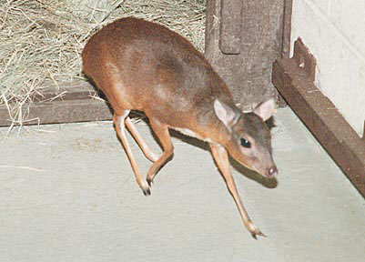
This animal, one of the three or perhaps more species of "dwarf antelopes", has the distinction of being the smallest antelope extant. The name Royal Antelope is said to derive from the local Liberian designation as being the "King of the Hares". Royal antelopes are West African animals and are rarely seen in zoos. They are very shy animals and thus difficult to photograph and perhaps do not serve as good exhibition specimens. Some of the related species of dwarf antelopes are more commonly seen in zoological gardens, especially the suni, Neotragus moschatus. Neotragus is often used synonymously with Nesotragus. The longevity of Royal antelopes is 6 years and 8 months, according to Jones (1993).
The details of the evolution of bovidae has been controversial. It is reviewed in some detail by Matthee & Robinson (1999). They suggested that the strictly African neotragini arose 12 MYA but then stated that "the assessment of evolutionary relationships in the dwarf antelope (Neotragini) has been troubled by many symplesiomorphic morphological characters, all possibly linked to their small size (Gentry, 1992)". They studied these controversies by analyzing the mitochondrial cytochrome b gene. It should be pointed out, however, that they did not have material from the Royal antelope available; it was restricted to suni, oribi, grysbok, steenbok, dik-diks and klipspringer. Of this group, only klipspringer and suni failed to follow a monophylogenetic grouping. These anomalies are discussed at some length and will require further work for resolution. Chromosomally, these animals also present some challenges with their cryptic chromosomal variation and with chromosome numbers ranging from 2n=30 in the steenbok to 2n=60 in oribi and klipspringer. Much more study is required before clarity of species designation is had.
Royal antelope female at San Diego Zoo.
2) General Gestational Data
Our pregnant female antelope came from Ghana and weighed 2.45 kg before giving birth. We did not weigh the fragile, single neonate but believe that it could not have been more than 300 g. No reference to twins was found in the literature, although oribi have had twins (Kellas, 1966). The length of gestation of Royal antelopes has not been described. That of the slightly larger suni is 180 days (Izard & Umfleet, 1971). They also reported that sexual maturity is reached at 6 months.
Adult female antelope with offspring.

3) Implantation
This has not been observed and can only be inferred from Kellas' paper on oribi which may be similar.
4) General Characterization of the Placenta
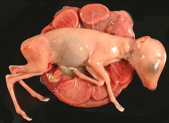
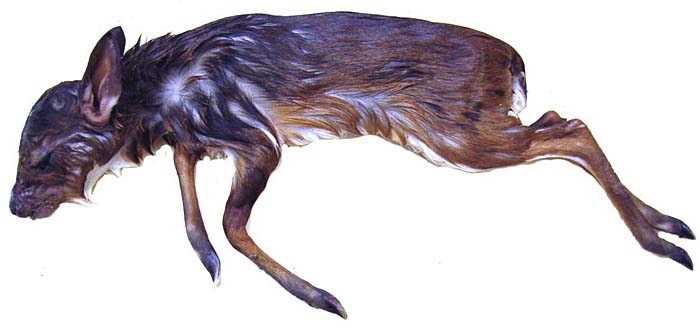
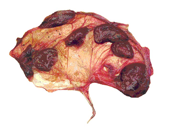
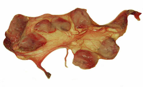
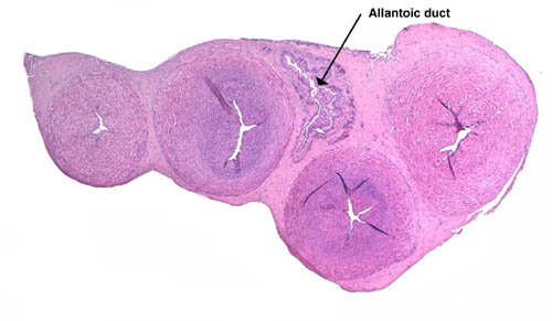
The placenta of this species has never been described, however, that of a related species, the oribi (Ourebia ourebi) has had a detailed description by Kellas (1966). The findings are similar, except that I did not find four but rather only two rows of cotyledons in the Royal antelope specimen. I have had the opportunity of examining only one placenta from an animal that came pregnant to our zoo and delivered a live infant. It had a polycotyledonary, epithelio-chorial placenta with only eight cotyledons, lined up in two rows. The placental specimen weighed 21.3 g, measured 17 cm in length, and the flat cotyledons were 3 cm in greatest width. They were only 0.2 cm in thickness. The thin, centrally inserted umbilical cord was 4 cm long and without spirals. A second placenta became available in May, 2004. It had essentially identical characteristics. It also possessed only 8 flat cotyledons ranging from 5.3 to 2.5 cm in diameters and had a 2 cm cord attached. A third placenta shown below had seven cotyledons and otherwise much the same morphology. It weighed 37.6 g. A third placenta became available in August 2005. It weighed 27 g and had 6 relatively large, flat cotyledons. The umbilical cord was 5 cm long. In February of 2007 one dam died with a fractured leg whilst pregnant with a 105.4 g female fetus. It had a 14 cm crown-rump length, weighing 105.4 g and the placenta had 9 cotyledons measuring from 2 to 4 cm in size and 0.4 cm in thickness. The umbilical cord was 9 cm long and 0.9 cm thick; its surface was studded with foci of squamous metaplasia. A photograph of that gestation is shown next. In January 2007 another term delivery occurred with a placenta that had 8 cotyledons and had an umbilical cord of 9 cm in length. And at the end of February 2007 another neonatal death occurred with a 170 g female neonate, a 30 g placenta that had 7 cotyledons and a 5 cm long umbilical cord.
Midgestational pregnancy with nine cotyledons.
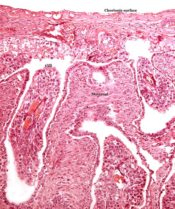
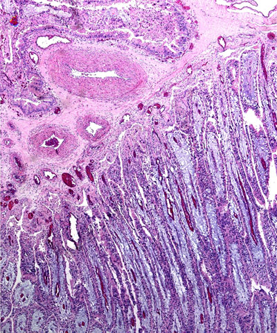
Implanted immature cotyledon.

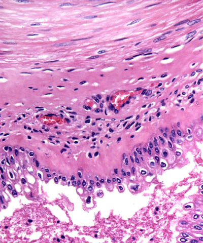
Surface of the immature placenta.
Base of immature cotyledon.
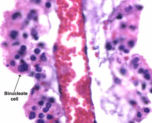
Villus (inside) with binucleate cell at arrow.

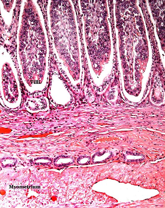
Implantation site between cotyledons with myometrium below.
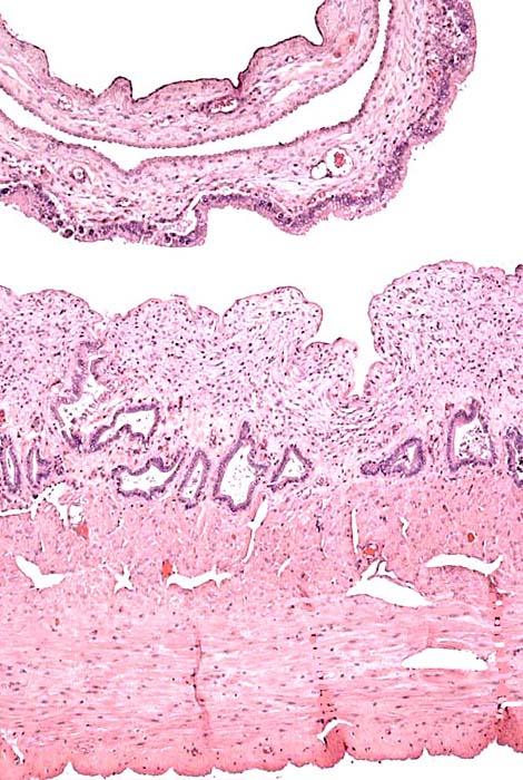
Free membranes of the immature placenta.

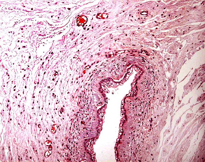
Allantoic duct in umbilical cord of immature placenta.
Female neonate, 170 g.
30 g placenta with 7 cotyledons of previous fetus.
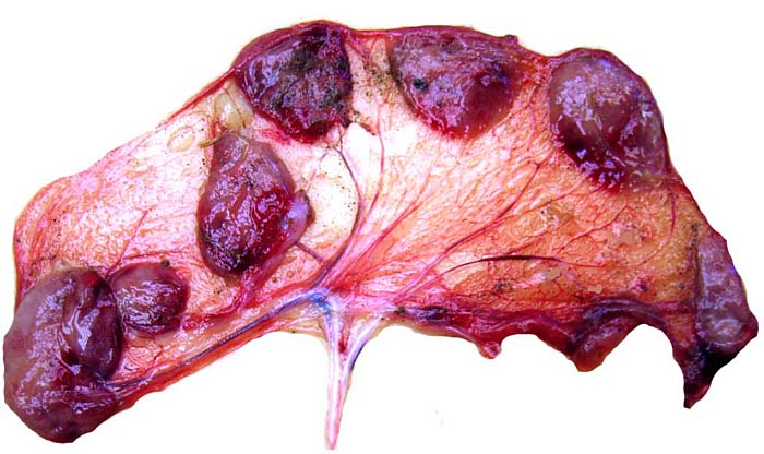
Third placenta from Royal antelope.
The villi were straight and little-branched. Their surface was covered with a single layer of cellular trophoblast and numerous binucleate cells. The autolytic process had caused some accumulation of fluid in the villi; otherwise they were similar to those described for oribi.
Macroscopic appearance of Royal Antelope placenta with tiny umbilical cord in the center.
Typical villous placenta that shows some degree of autolysis in the blue connective tissue of the villi. Chorionic plate with fetal vessels above.
5) Details of fetal/maternal barrier
There is a single layer of cuboidal trophoblast on the surface of the villi, with a much taller trophoblast on the chorionic plate that extends between the cotyledons. A moderate number of characteristic binucleate cells (producing relaxin) was found along the villous surfaces.
Section of one villus with mild autolysis. Fetal vessel in center, disorganized (autolyzed) trophoblast contains a moderate number of binucleated cells.
6) Umbilical cord
The umbilical cord was centrally inserted and 4 cm long without spirals. It was 0.3 cm in thickness. There were two arteries and two veins, as well as a large allantoic duct. The surface was smooth. Two additional cord lengths were 2 and 5 cm.
Section of thin umbilical cord with large allantoic duct at arrow. The blood vessels are conspicuously confined to the periphery of the duct.
Higher power of the wall of the allantoic duct with its adjacent blood vessels. Debris inside the duct.
7) Uteroplacental circulation
This has not been studied.
8) Extraplacental membranes
The amnion is rather usual, with a thin, squamous amnionic epithelium and lacking blood vessels. The allantoic sac was very small and empty. Its wall had a large number of blood vessels and it was clad also by a thin epithelium. No hippomanes were found. There was, however, debris in the allantoic duct and in the allantoic sac. There was no decidua capsularis on the membranes.
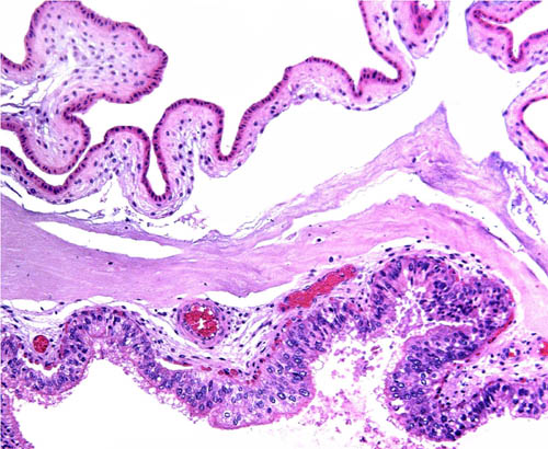
Amnion above, allantois with allantoic blood vessels below.

Membranes between the cotyledons. Amnion is above, the trophoblastic layer overlying the areolae below. Note the cylindrical trophoblast and debris over areolae.
9) Trophoblast external to barrier
No extravillous trophoblast is likely to be present, but no implanted placenta has been described.
10) Endometrium
This is unknown, as no term placenta has been described that was attached to the uterus.
11) Various features
Except for the extremely small size, no unusual features were observed.
12) Endocrinology
No studies are known to me.
13) Genetics
The chromosome number of neotragini is complex. Kingswood et al. (1998) studied phenotypically similar specimens of the suni (Neotragus moschatus) from several zoological gardens. They found chromosome numbers varying between 2n=52 and 2n=56. This raised the question whether the specimens belonged to different subspecies or, whether these chromosomally divergent karyotypes should be assigned to different species, albeit look-alikes. Studies from the wild populations have not been undertaken to clarify this question. The results just cited also indicate that perhaps intermediate forms (?hybrids) of animals with this "cryptic chromosomal variation" are subfertile. Similar problems have been identified in dik-diks and in a few other African gazelles. True, known interspecific hybrids have not been described. The chromosome number of Royal antelopes has not yet been determined. Our cultures are in progress and, now finished, are added at this place. The interesting result of these cultures is that the chromosome number is 2n=36, with numerous acrocentrics. This raises interesting questions that we are currently exploring.
The genetic study of the mitochondrial cytochrome b gene gene (Matthee & Robinson, 1999) was referred to at the beginning of the chapter.
Male and female karyotypes of Royal antelopes from the San Diego Zoo.

14) Immunology
No studies are known to me.
15) Pathological features
Izard & Umfleet (1971) reported infection with Haemonchus contortus and strongylodiasis in their suni antelopes.
16) Physiologic data
No studies are known to me.
17) Other resources
Cell lines of the suni are available from the study of Kingswood et al. (1998). The lines from the Royal antelope will also become available. They can then be obtained by contacting Dr. Oliver Ryder at: [email protected].
18) Other remarks - What additional Information is needed?
Implanted placentas are needed to fill in the gaps of knowledge. The exact length of gestation remains also unknown.
Acknowledgement
The animal photograph in this chapter comes from Ron Garrison at the Zoological Society of San Diego. I appreciate also very much the help of the pathologists at the San Diego Zoo.
Gentry, A.W.: The subfamilies and tribes of the family Bovidae. Mamm. Rev. 22:1-22, 1992.
Izard, J. and Umfleet, K.: Notes on the care and breeding of the suni Nesotragus moschatus at Dallas Zoo. Int. Zoo Ybk.11:129, 1971.
Jones, M.L.: Longevity of ungulates in captivity. Int. Zoo Ybk. 32:159-169, 1993.
...About
How to reference this publication (Harvard system)?
Affiliation of the authors at the time of publication
Department of Reproductive Medicine and Pathology, School of Medecine, University of California, San Diego, CA, USA.

Comments (0)
Ask the author
0 comments