
List of Definitions: The Common Dental Disorders in Donkeys
Author(s):
Updated:
FEB 09, 2021
Languages:
Read
The most commonly found dental disorders in donkeys have been listed here, along with a short description of the condition. The accompanying images have been selected to illustrate the definition and have been taken from the extensive library of images held by The Donkey Sanctuary and from those belonging to the contributors of this book.
Apical infection
Extension of pulpar disease through the apical foramen into the peri-apical (apical) periodontal tissues, with infections usually spreading around the apex, causing clinical changes to the alveoli and supporting bones.
Bacterial infection may follow exposure of the pulp to the oral environment.This may occur due to complicated fractures, but also in more severe cases of caries, where all dental components are affected, leading to apical infection. Bacteria may also reach the pulp cavity via periodontal spread but mainly via anachoresis (blood-born bacterial infection), decreasing the production of secondary dentine by the odontoblasts, and leading in time to pulp exposure due to the continuous dental wear process.
This last source of bacterial infection appears to be the most common cause of Cheek Teeth (CT) apical infections.
Vertical impaction/overcrowding or retained deciduous teeth may also contribute to this disorder.
The clinical signs caused by apical infections are directly related with the site and age of the infected tooth, and the duration and the extent of the infection.
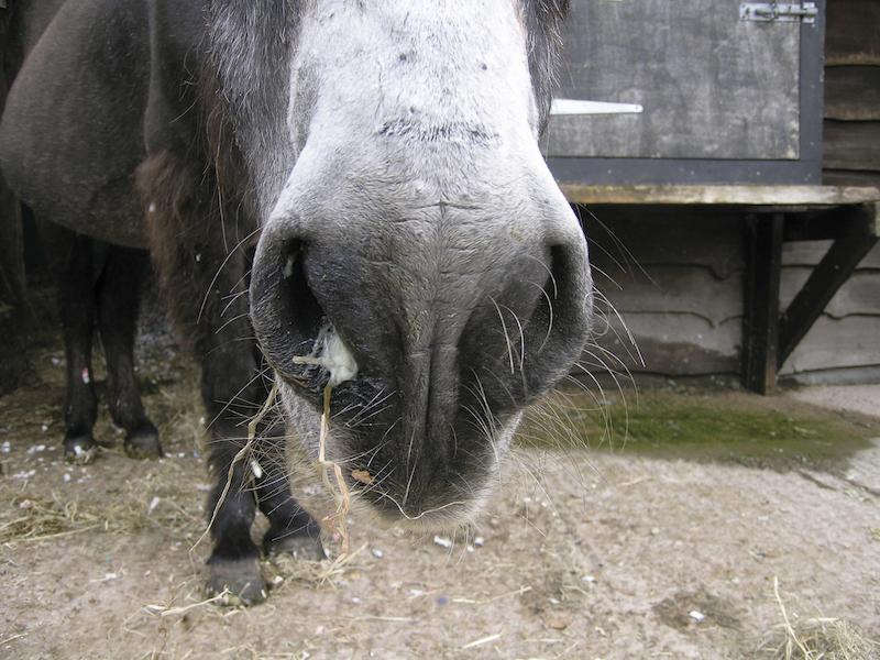
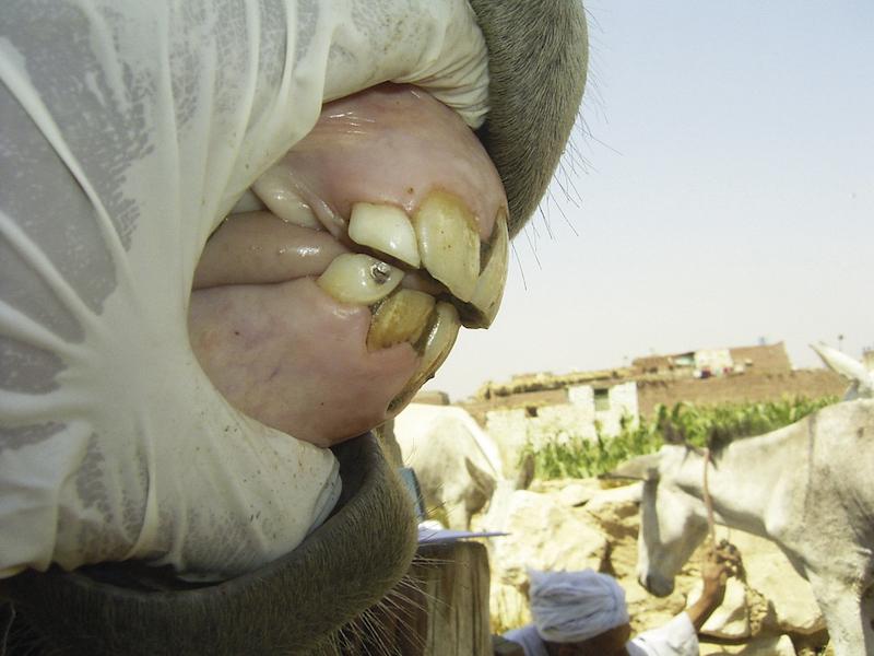
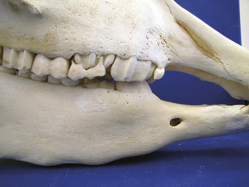
Unilateral mucopurulent nasal discharge, due a secondary sinusitis caused by an apical infection in 109.
Caries
Caries is described as the progressive dissolution of the calcified components of a tooth as a result of bacterial fermentation of carbohydrates. In equids, dental caries is a condition which is typically observed within the infundibulum (infundibular caries) and/or at the peripheral aspects of the tooth (peripheral caries lesions).
Infundibular lesions are graded according to severity:
1. only the cementum
2. cementum and adjacent enamel
3. cementum, enamel and dentine
4. affecting the integrity of the tooth (the most severe cases).
Peripheral lesions are graded according to severity:
1.1. only the cementum is affected
1.2. only the cementum is affected; a greater degree of cementum is affected to the extent that there are areas of complete loss. The enamel is exposed, but unaffected.
2. cementum and underlying enamel is affected
3. cementum, enamel and dentine are affected.
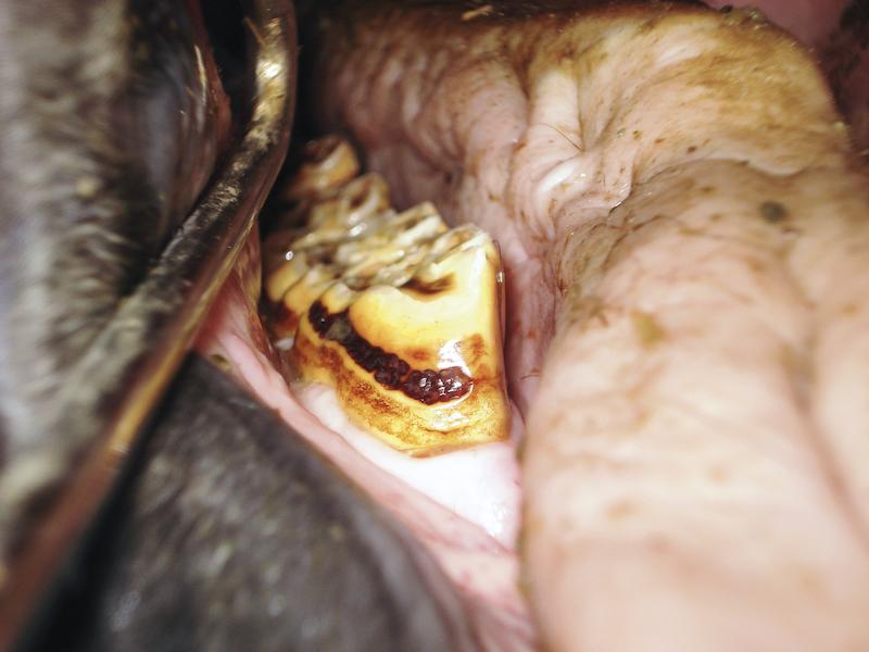
Infundibular caries in 3 and 402. Note the enlargement of the infundibulum in the affected tooth.
406 with supragingival grade 1.1 peripheral caries.

Coalesced infundibular owing to rostral and caudal infundibular caries.
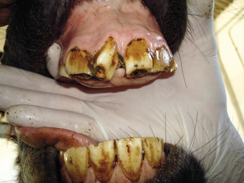
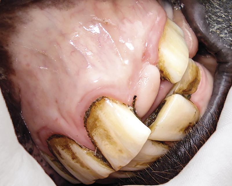
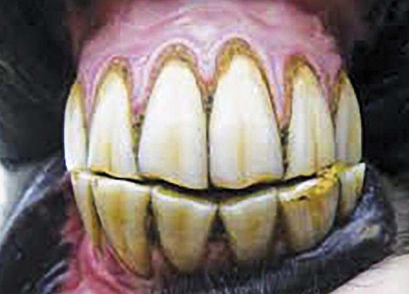
Calculus formation
A result of the mineralisation of the dental bacterial plaque, often found in cases with significant malocclusions at dentition local to salivary ducts.
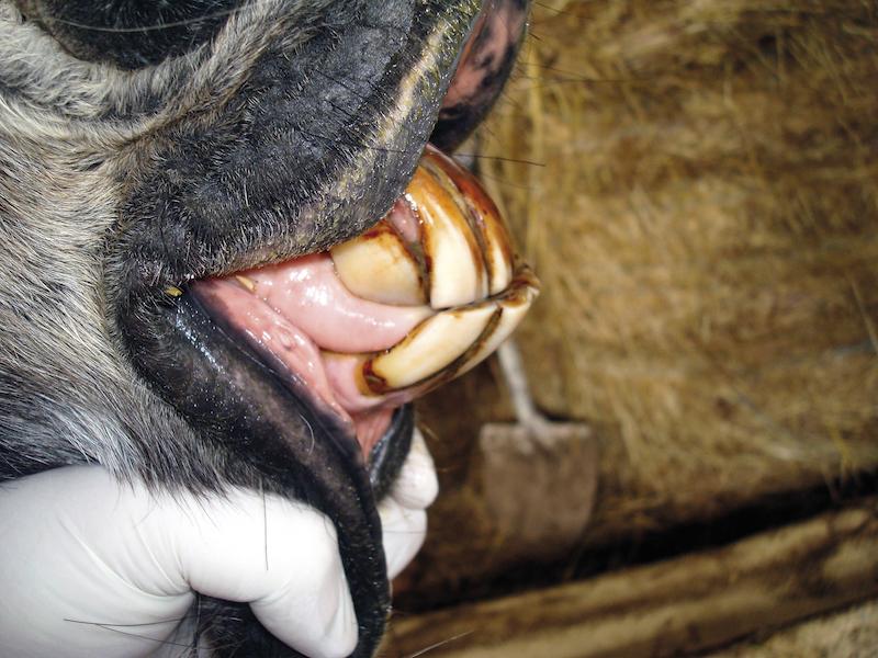
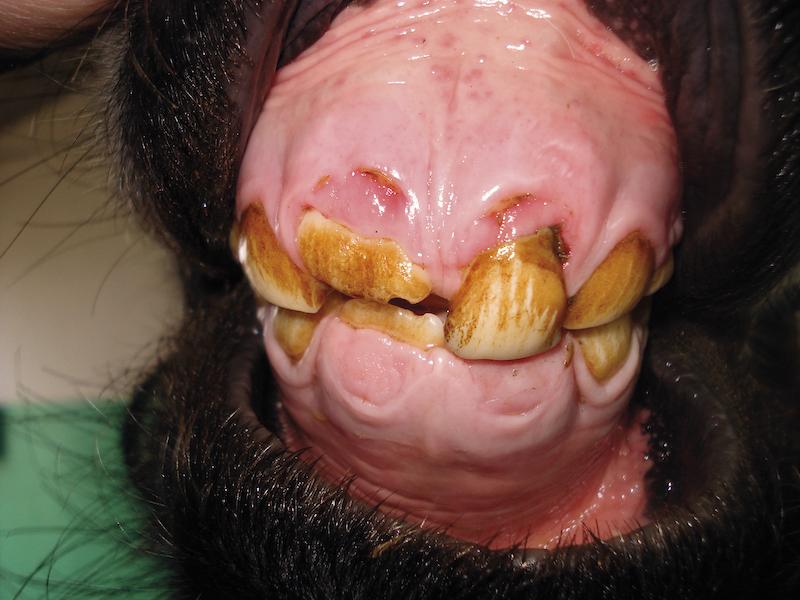
Presence of calculus in the third arcade, affecting the mandibular incisors and the canine. In humans and small animals, it's proven that surface roughness alone doesn’t initiate gingivitis, but that plaque retention causes periodontal disease (PDD); removal of plaque from calculus deposits will often result in healing of periodontal lesions.

Calculus deposition at the 108 and 109.
Craniofacial abnormalities
Uneven upper and lower incisor occlusion, as a result of growth abnormalities in the craniofacial bones. Cheek teeth may also be affected in these situations, mainly 06s and 11s.
This includes:
Overjet
Occlusal aspects of the maxillary incisors lie rostral to the occlusal aspect of the mandibular incisors, with contact between teeth.
Underjet in an adult donkey, with inversion of the normal angle in the incisors’ occlusal surface, changing from a 10° to 15° angle in a dorso-caudal to rostro-ventral inclination, to a ventro-caudal to dorso-rostral sense, promoting a slight overgrowth in the labial aspect of the mandibular incisor occlusal surface and in the palatal aspect of the maxillary occlusal surface.
Overjet with overbite
Maxillary incisors lying rostral and in front, without any occlusal contact between upper and lower incisor arcades. This is also known as Brachygnathism.

Newborn foal presenting extreme case of overjet with overbite. Such extreme situations may compromise the suckling capacity of the offspring, threatening their ability to survive.
Underjet
Occlusal aspects of the mandibular incisors lie rostral to the occlusal aspect of the maxillary incisors, with contact between teeth.
Underjet in an adult donkey, with inversion of the normal angle in the incisors’ occlusal surface, changing from a 10° to 15° angle in a dorso-caudal to rostro-ventral inclination, to a ventro-caudal to dorso-rostral sense, promoting a slight overgrowth in the labial aspect of the mandibular incisor occlusal surface and in the palatal aspect of the maxillary occlusal surface.
Underjet with underbite
Mandibular incisors lying rostral and in front, without any occlusal contact between upper and lower incisor arcades. This is also termed Prognathism.

Extreme case of underjet with underbite.
Variable degrees of lateral premaxillary (incisive) and maxillary bone deviation; which are known as campylorrhinus lateralis or ‘Wry nose’.

Three year-old donkey with wry nose, with the maxilla deviated more than 50° angle when compared with the mandible.
Diastemata
An abnormal increase of the interproximal space, with or without food pocketing, and includes valve and open diastemata.
Distal positioning of the deciduous mandibular corner incisors, resulting open diastemata formation.

Post-mortem image of multiple bilateral open diastemata, affecting most of the mandibular interproximal space.

Senile diastemata (acquired) affecting interproximal space in the mandibular and maxillary incisor arch in a geriatric donkey.
Post-mortem image of a diastema between 410 and 411. Note the presence of a foreign body in the diastema, causing severe lacerations of the tongue and buccal mucosa.
Dental displacements
Variation of the physiological position within the incisor arch or CT row. Displacements may be devel- opmental or acquired.
In the incisors, displacements can be vestibular, lingual/palatal or distal, according to the direction of deviation inside the mouth.
In the CT, displacements can be in a vestibular, lingual/palatal, distal or rostral sense.
Teeth can also rotate on their own axis, within the incisor arch or CT row.
Developmental axial rotation of 90° affecting 101. Note the presence of open diastema between 101 and 201, without periodontal disease.

Severely displaced 308.
Developmental axial rotation of 45° affecting 208, observed in a donkey skull.
Dysplastic teeth
Developmental disturbances in the correct gross form of the teeth.
These include dilacerations (abnormal distortion of the teeth), disturbances in the size and shape, distortion and concrescence, with roots of adjacent teeth joined by cementum.
Donkey with mandibular dysplastic incisors and developmental hypodontia.
Intra-oral view of the mandibular incisors, showing dysplastic teeth and developmental hypodontia. The intra-oral bisecting angle technique was used to allow a correct diagnosis.

Dysplastic 108 and 109 in a young donkey.
Eruption cysts
Enlarged, focal swellings, beneath young permanent premolars, in the developing apex region, palpable in the mandible and maxillae regions.
Skull of a 3 year-old donkey, showing an irregular mandible due to the presence of eruption cysts beneath the premolars. Note the presence of the 507 cap.
Fractures
Typically the result of pathological or traumatic injury of the teeth, affecting part or all of its components:
Uncomplicated fracture
Only the external components of the teeth (cementum, enamel and sometimes, the dentine) are affected.
Complicated fractures
The pulp cavity is directly or indirectly exposed.
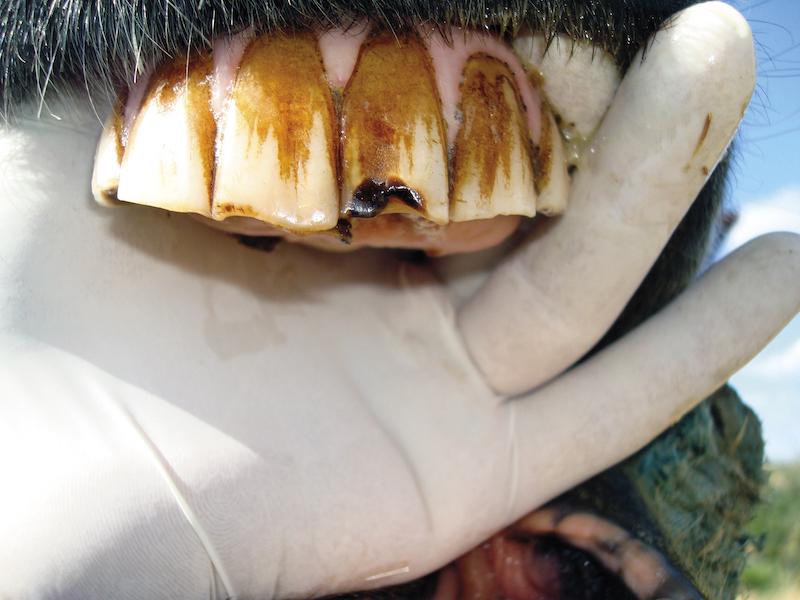
Uncomplicated fracture in 201. These small fractures, located in the transition area between the occlusal surface and the labial aspect of the teeth, can be a predisposing factor to the development of peripheral caries.
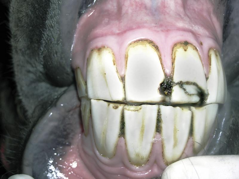
Typical traumatic incisor fracture
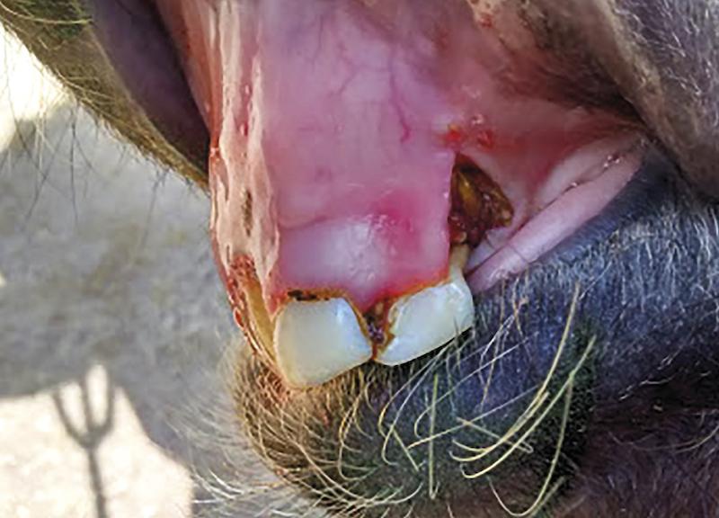
Donkey with an avulsion of the deciduous incisors and premaxilla.

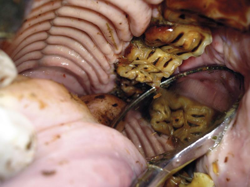
Oroscopic view of a complicated sagittal fracture in a CT.
Hypodontia
A reduced number of teeth. This can be developmental, as a result of an inappropriate differentiation of dental lamina and tooth germs (agenesia), or may occur during the animal’s lifetime, due to trauma, age or secondary to other dental disorders.
Developmental hypodontia in a donkey. Note the presence of all definitive incisors, with 202 missing.

Geriatric donkey with severe acquired hypodontia, as a result of age and dental disorders.
Overgrowths
Abnormalities of the occlusal surface are seen in both incisors and CT. In the incisors it may occur as:
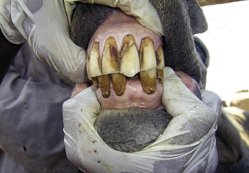
Ventral and dorsal convex curvatures, diagonal or irregular variations in the occlusal line of the upper and lower incisors.
Ventral curvature of the incisors.

Acquired dorsal convex curvature in the occlusal surface of incisors in an adult donkey.
Diagonal incisor malocclusion in a geriatric donkey.
Extreme case of irregular malocclusion in a donkey, probably as a consequence of an old fracture affecting the mandibular symphysis.
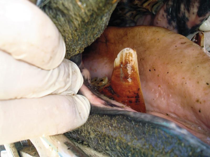
Incisor overgrowth
Incisors that have not been exposed to normal wear, usually secondary to craniofacial abnormalities, or due to disorders affecting the opposing teeth (fractures, displacements, loss or excessive wear).
Overgrowth of 201, due to the absence of the ipsilateral opposite tooth (301).
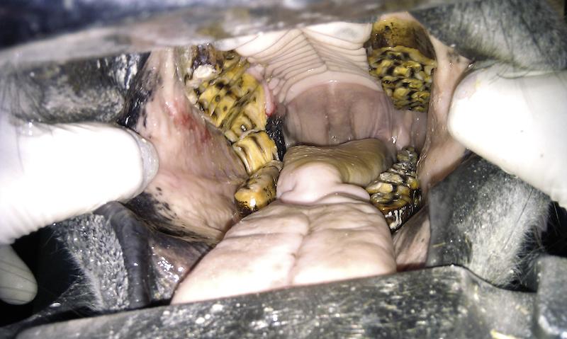
In the cheek teeth these are described as any variations affecting the normal wear process/attrition between ipsilateral opposing teeth, leading to a restricted mandibular movement and increasing the possibility of soft tissue damage. These are seen as:
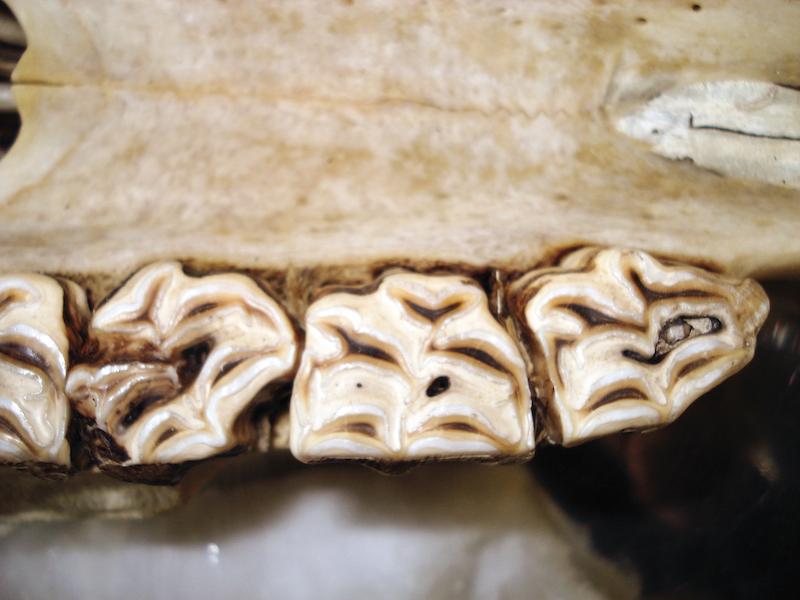
Cheek teeth enamel overgrowths
Development of protruding enamel overgrowths, usually in the lingual aspect of the mandibular CT and vestibular aspect of maxillary CT, due to the presence of anisognathism and the angled conformation of the CT occlusal surface.

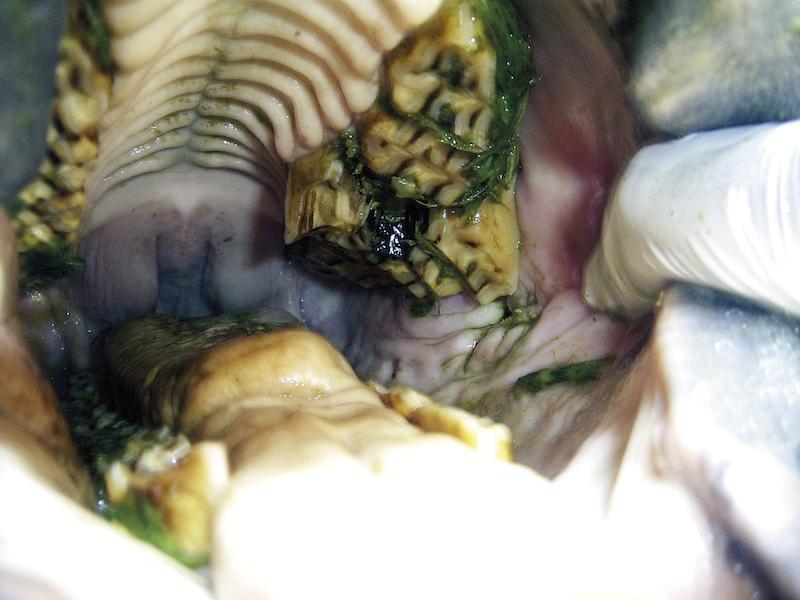
Post-mortem CT enamel overgrowths, affecting mainly the molars (09 to 11s).
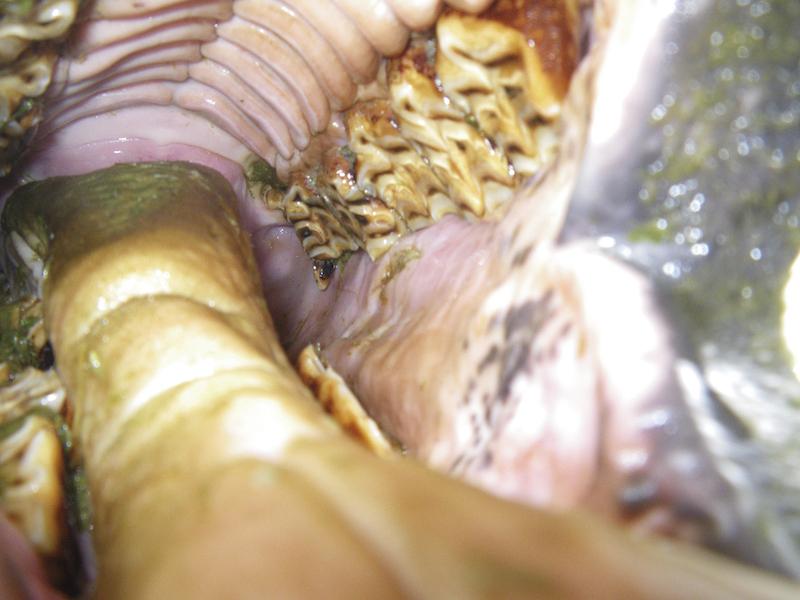
Cheek teeth overgrowths
Complete or partial CT surface that has not been exposed to normal wear, usually secondary to cranio-facial abnormalities (focal overgrowths, affecting the 06s and 11s), or due to disorders affecting the opposing teeth (dysplasia, displacements, fractures, loss or excessive wear).
Complete CT overgrowth affecting 109 and 110 due to dysplasia of the ipsilateral opposite teeth.
Bilateral focal overgrowth in the mandibular 06s, in a donkey with underjet with underbite.
Wave mouth
Undulating appearance of the occlusal surface, in a rostro-caudal sense.

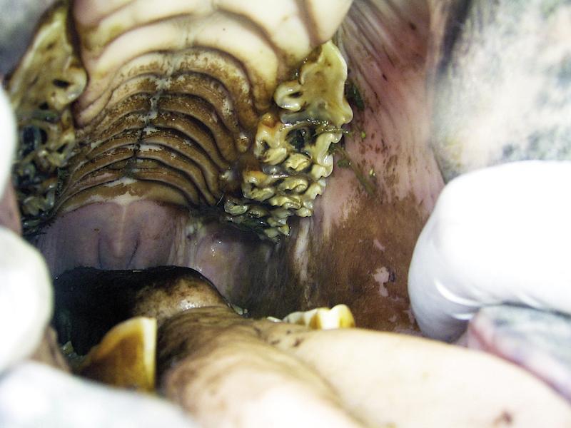
Wave mouth affecting the 2nd and 3rd cheek teeth rows.
Shear mouth
The presence of a steeper angle in the occlusal surface of the CT row (usually >45°), in the buccal-palatal sense.
Advanced case of shear mouth.
Smooth mouth
Reduction or complete loss of enamel ridges, but defined as worn teeth or senile excavation if only a limited number of teeth are affected.
Geriatric donkey with senile wear mainly affecting the maxillary 06s.
Periodontal disease
A pathological process affecting the periodontum, ranging from gingivitis without attachment loss (stage 1) to severe, deep periodontal pocketing (stages 2, 3 and 4 with < 25%, < 50% and more than 50% of attachment loss, respectively).

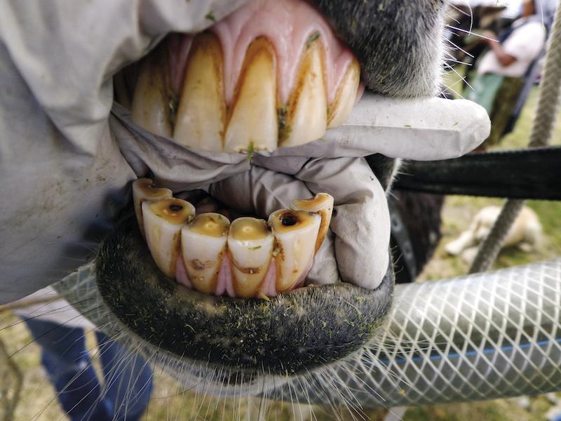
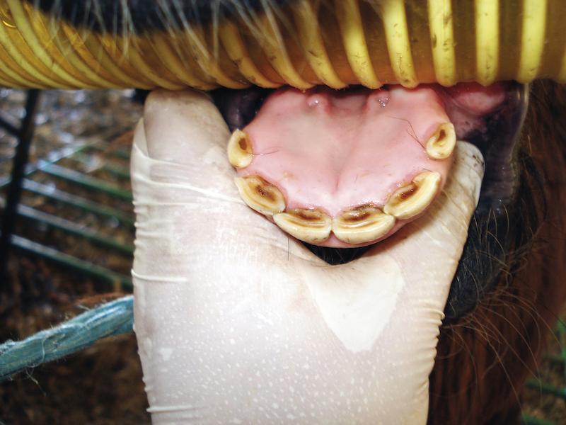
Gingivitis (periodontal disease stage 1) due to the presence of calculus in the lower incisors.

Extreme case of periodontal disease stage 4, affecting all the maxillary incisors, with severe calculus accumulation and peripheral caries in 201 (stage 4).

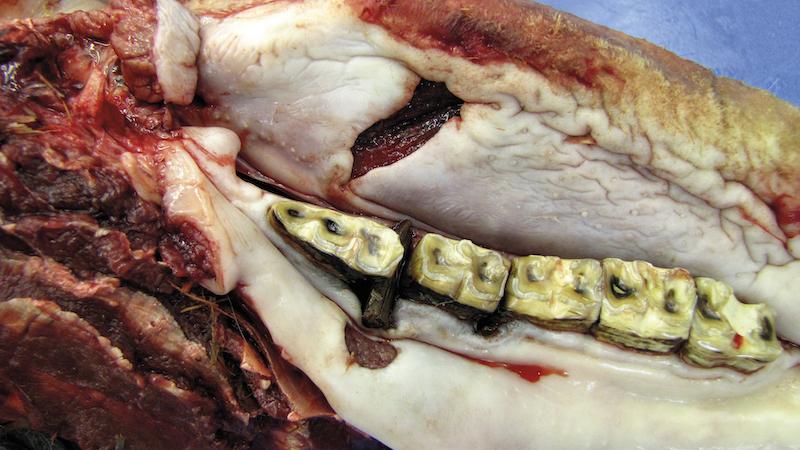
Post-mortem image of a CT diastema, affecting 408 and 409. Note the deep periodontal pocket and gingival recession around the affected area.
Retained deciduous teeth
The presence of deciduous teeth beyond their normal time of shedding, affecting the normal eruption process of the permanent dentition.
Donkeys commonly exchange the deciduous teeth 6 to 8 months later than recorded in horses and ponies.
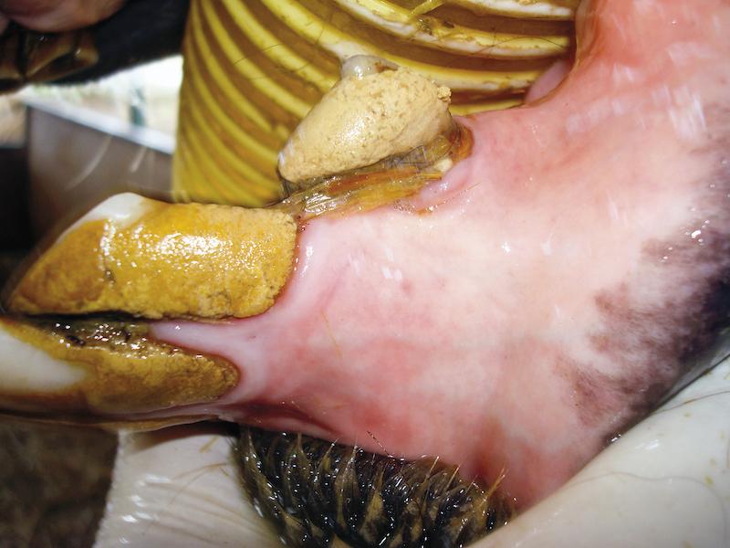
Retained 601. Note the presence of retained root fragments both in 501 and 601. These fragments act in many cases as a source of gingival irritation and should also be extracted, by elevating the gum in a conservative way, avoiding unnecessary lacerations and trauma.

Bilateral caps at 506 and 606. Only caps being digitally loose, having partial loss of the crown, clear and palpable demarcation between deciduous and definitive teeth at the gum level should be considered retained and therefore extracted.
See chapter 1 for information on eruption of deciduous teeth.
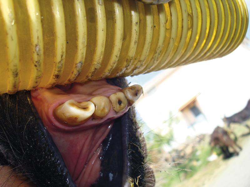
Supernumerary teeth
The presence of teeth in excess of the normal dental formula, as a result of an inappropriate differentiation
of dental germinal tissue during gestational development. External trauma may be an initiating factor and teeth germs may be affected;

Unilateral unopposed maxillary distomolar CT supernumerary 111, presenting clear overgrowth/lack of wear.
Supernumerary teeth are classified as:
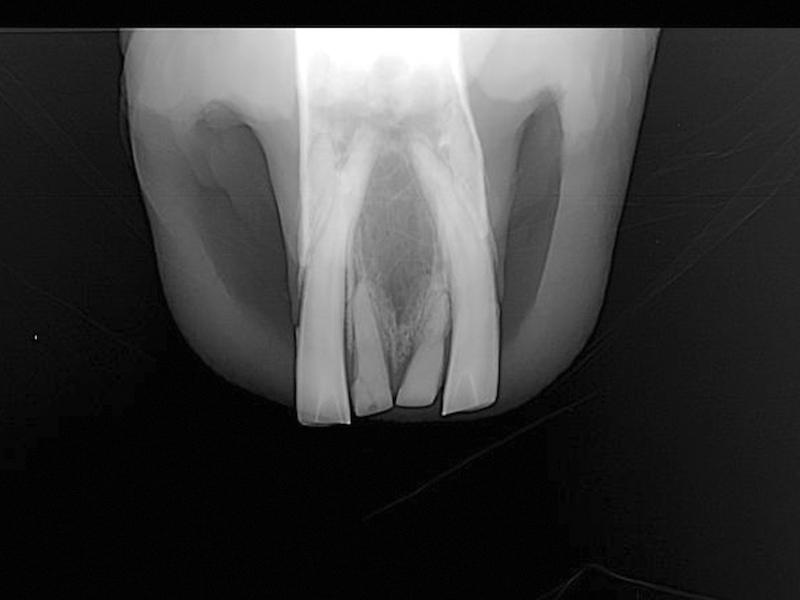
Supplemental/eumorphic teeth
Presenting normal crown and root morphology.
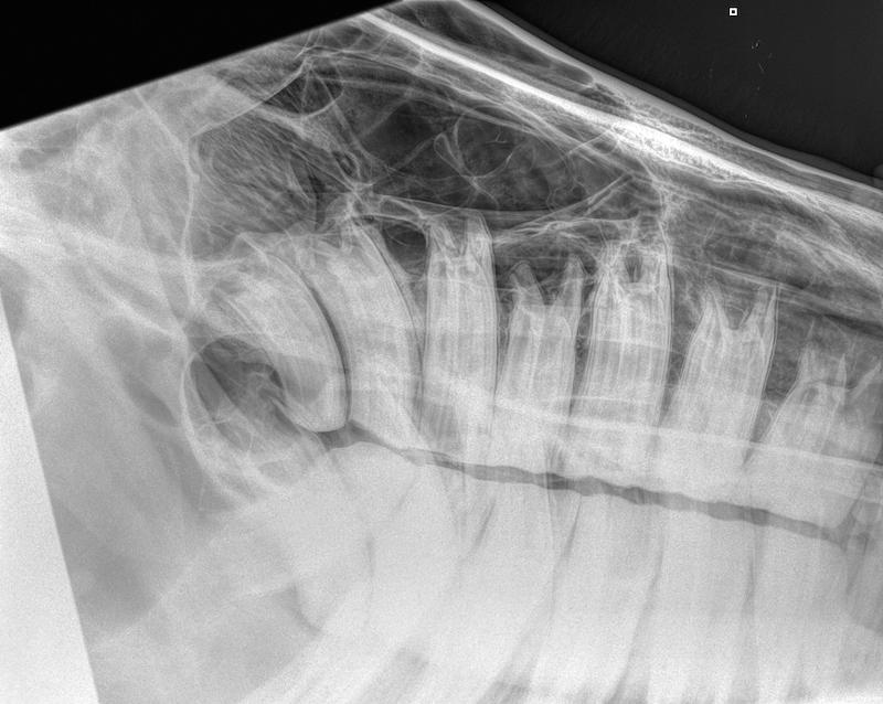
Radiographic latero 30° dorso- lateroventral oblique projection of the left maxillary arcade in a donkey, showing the presence of a supernumerary eumorphic 212.
Supernumerary 208 palatally adjacent to CT row, causing the buccal displacement of 208. Note the diastemata between both 208, as well as with 207 and 209, with periodontal disease development.
Rudimentary/dysmorphic teeth
Presenting abnormal size and shape:
-
haplodont (conical crown with a single root).
-
tuberculate (several tubercles with deep indentations on the occlusal surface).
Germinate/Connate teeth
Germinate teeth are a result of division of a single bud or connate teeth are created by fusion of two buds via dentine, and concrescence created by fusion at the cementum.
Intra-oral view of a connate 208, with 3 infundibulum and 9 pulp horns present in the tooth surface.

About
How to reference this publication (Harvard system)?
(2021) “List of Definitions: The Common Dental Disorders in Donkeys”, The Clinical Companion of Donkey Dentistry. Available at: https://www.ivis.org/library/clinical-companion-of-donkey-dentistry/list-of-definitions-common-dental-disorders-donkeys (Accessed: 19 May 2024).
Affiliation of the authors at the time of publication
Sidmouth, Devon, EX10 0NU
Author(s)
Copyright Statement
© All text and images in this publication are copyright protected and cannot be reproduced or copied in any way.Buy this book
Buy this book
The Clinical Companion of Donkey Dentistry is an easy reference book for professionals produced as part of a series of specialist books that will compliment The Clinical Companion of the Donkey. It enables us to share our vast knowledge and experience to improve the health and welfare of donkeys globally.
Following on from the publication of The Clinical Companion of the Donkey, we plan to produce a series of in-depth specialist handbooks which will complement the handbook.
This book is intended as a guide to the anatomical features of the head and oral cavity of the donkey, to offer a greater understanding of the oral and dental disorders that may affect these animals throughout their life, and how to correctly examine, diagnose, prevent and/or treat pathological situations.
Dentistry is the first topic to be published in this series, and we consider it to be an area which is extremely important to the health and welfare of donkeys globally, while being misunderstood and undervalued by many communities.
This book allows us to share our vast knowledge and experience in donkey specific dentistry and has been produced as an easy reference and well-illustrated book, which we believe will not only increase awareness, but also the confidence of professionals in carrying out dental care and treatment in donkeys.
Comments (0)
Ask the author
0 comments