Developmental Orthopedic Diseases Associated with Nutritional Excesses
Read
3. Developmental Orthopedic Diseases Associated with Nutritional Excesses
Osteochondrosis
Osteochondrosis is a disturbance of endochondral ossification, characterized by an abnormal maturation of chondrocytes and therefore a delay in cartilage mineralization (Figure 7 and Figure 8). If the disturbance in endochondral ossification occurs in articular cartilage, osteochondritis dissecans (OCD) may be a consequence. In OCD, part of the articular cartilage becomes detached and may be fragmented, mineralized or even ossified together with inflammation of both the joint and the endochondral bone in the area of the cartilage lesion.
Figure 7. Osteochondrosis lesion. Osteochondrosis is the result of an anomaly in the development of growing cartilage: the ossification process is altered and we see cartilage retention and thickening. (The arrow points to a zone of abnormally thick cartilage.) Osteochondrosis may develop into osteochondritis dissecans when a fragment of cartilage is freed in the joint. Osteochondrosis is stimulated by a chronic excess of calcium in the diet.

The disturbance in endochondral ossification can also occur in growth plate cartilage, with irregular growth plates, enlarged cartilage cores and decreased growth in length as a consequence. In addition, the disturbance in endochondral ossification can manifest in a delay in ossification of the secondary ossification centers. Disturbances in growth plates which lead to clinical manifestations of osteochondrosis (including bilateral radius curvus syndrome or rear leg exorotation) are seen predominately in giant breed dogs. Detachment of the anconeal process of the ulna or the supraglenoid process of the scapula can also occur.
Figure 8. Normal (left) and abnormal (right) process of endochondral ossification in the growth plates and growing articular cartilage.

Diagnosis
Dogs with osteochondrosis, without detachment of cartilage, will not be clinical. However with OCD, dogs may exhibit lameness, pain upon hyperextension and flexion of the affected joint and joint effusion. The joints most commonly affected are shoulder, elbow, stifle and hock. Dogs will also not be clinical in cases of osteochondrosis in the growth plate when the retained cartilage core is small or temporary. In longitudinal radiological studies of the distal growth plate of the ulna of Great Danes, a slight flattening or indentation could be seen at five months of age. However, when a severe flattening of the metaphyseal area develops, or a deep cartilage core can be seen, impaired growth in length of the radius and ulna can be expected (Figure 9). This can result in radius curvus syndrome (short ulna, curved radius and valgus deformation of the feet).
Figure 9. A mild retained cartilage core may disappear spontaneously.

With clinically suspected OCD, a thorough clinical and radiographic investigation will suffice in most cases. Radiographs may reveal subchondral sclerosis bordering an indentation of the articular surface. Some cases may require additional diagnostics such as arthrocentesis, contrast arthrogram, arthroscopy, other imaging techniques and/or exploratory surgery of the joint for definitive diagnosis.
Epidemiology
Osteochondrosis is primarily seen in large breeds of dogs (i.e., more than 25 kg adult body weight), and more frequently in male and fast growing female dogs. The disease is seen in a variety of breeds with Great Dane, Labrador, Golden Retriever, Newfoundland and Rottweiler being the breeds at highest risk (Milton, 1983; Slater et al., 1991; Van Bree, 1991).
Osteochondrosis appears to have a predisposition for specific locations in each breed.
OCD lesions are seen most commonly in:
- The shoulder and stifle joints of Great Danes
- The shoulder, elbow and hock joints of Labradors and Golden Retrievers
- The elbow joints of Newfoundlands
- The shoulder and hock joints of Rottweilers (Slater et al., 1991).
In one study, 66% of the dogs with OCD had more than one joint affected; in 5% three joints were affected (Slater et al., 1991). Although histologically osteochondrosis can be diagnosed also in non-weight bearing growth plates like that of the rib, microtrauma may play a significant role in causing the fissures as can be concluded from the fact that osteochondrosis dissecans is mostly seen on convex, weight bearing areas.
Pathophysiology
Osteochondrosis is a multifactorial disease common in large breeds in which heredity and nutrition play a significant role. A variety of studies have been performed to elucidate the role of nutrition in the manifestation of osteochondrosis (Table 2). These studies support the conclusion that chronic intake of excess energy or of a food enriched with calcium, with or without changes in other nutrients, plays a significant role in the development and manifestation of osteochondrosis in dogs of large breeds. Minor changes of endochondral ossification, without clinical significance, have also been demonstrated in miniature Poodles raised on a food with high calcium content. Please refer to sections: Overnutrition in Growth and Excess Calcium Intake; for further information.
Therapy
Dietary correction at an early stage may positively influence the spontaneous resolution of disturbed endochondral ossification (Voorhout & Hazewinkel, 1987a). Osteochondrosis in cartilage and growth plates could disappear, but dietary modification will not normalize cases of OCD in which there is severe detached cartilage, or where a more severe curvature of the radius exists (Olsson, 1982). Surgical correction will be indicated in most of these cases (Figure 10).
Figure 10. Arthroscopic removal of a cartilage flap from the humeral head.

Dietary correction entails a decrease in intake of energy, calcium and vitamins to the recommended levels for dogs. Fast growing puppies should be weighed and have their body condition score assessed frequently (every 2 - 4 weeks) to monitor their growth. If a puppy is growing too quickly or becoming overweight, its energy intake will need to be decreased without unbalancing the other nutrients. No pharmacological or other medications are known to support the nutritional management.
Osteochondrosis will not develop into OCD in all cases. Based on controlled studies where both shoulder joints were radiographed, it can be concluded that, although 45 - 65% of the dogs had radiographically detectable abnormal contours of the humeral head, only 3 - 5% were clinically affected on both sides (Van Bree, 1991). When detached, the period of lameness can be shortened as well as most probable the secondary changes of the joint minimized with surgical or arthroscopic treatment.
Elbow Dysplasia (ED)
ED can be separated into different disease entities including ununited anconeal process (UAP), fragmented coronoid process (FCP), osteochondritis dissecans (OCD) of the medial humeral condyle and incongruities of the elbow joint (INC).
Diagnosis
The typical age of the patient suffering from ED is 4 - 10 months of age, although there is an increasing frequency of dogs with elbow pain (without any signs of OA on radiographs) seen at more than 3 years of age. Upon examination in almost 50% of the cases the paw of the affected leg is externally rotated and slightly abducted. On palpation the elbow is often effusive. The range of motion (ROM) of the elbow joint can be decreased in advanced cases. Subtle crepitation can be appreciated at an early stage. In an UAP, there is crepitation and pain on firm hyperextension of the elbow joint. In FCP and/or OCD, crepitation and pain reaction can be evoked at prolonged hyperextension, especially when the radius and ulna are externally rotated at the same time (i.e., supination).
Diagnosis of ED can be confirmed by radiography. Bony union between the anconeal process and the olecranon should be complete at the age of 16 - 20 weeks (Sjöström et al., 1995). When a radiolucent area is present at an older age it is suggestive of an anconeal process which is not united, i.e., an UAP. This may be due to a partial or complete separation in the cartilage between the anconeal process and olecranon, which is preferentially demonstrated on a mediolateral flexed radiographic view. Sclerosis at the fracture site and osteophytes at the margins of the joint can be visible at a later stage. For grading OA of the elbow joint, see the web page of the International Elbow Working Group: www.iewg-vet.org.
OCD of the medial humeral condyle is best assessed on anterior posterior medial oblique (APMO) views (Voorhout & Hazewinkel, 1987b). In a small number of cases a calcified flap can be seen located near the indentation of the medial condyle. In FCP, the fragment can only be seen on high quality films when the coronoid process is displaced cranially (as in Bernese Mountain Dogs), whereas the cranial alignment of the ulna at the level of the medial coronoid can give an indication of fragmentation. Secondary signs such as osteophytes and sclerosis of the semilunar notch can help to confirm the clinical diagnosis. Small osteophytes are especially visible on mediolateral views of the flexed elbow at the dorsal margin of the anconeal process. Extended views help to visualize osteophytes at the radial head, and AP views demonstrate irregularities at the medial aspect of the humerus as well as the ulna (Figure 11).
Figure 11. Elbow joint of an 8-month-old Bernese Mountain Dog with severe osteoarthrosis (OA). (A) Notice osteophytes at the radial head, anconeal process and medial epicondyle and osteosclerosis in the semilunear notch. (B) The craniocaudal view shows that the radial head does not run parallel with the distal humerus indicating elbow incongruity as well as an indication of a fragmented coronoid process (FCP). The final diagnosis is: elbow incongruity and FCP with severe OA. (© HAW Hazewinkel).

Most entities of ED occur bilaterally in 30 - 70% of the cases, and therefore both elbow joints should be investigated, even in case of unilateral lameness. In case there are no radiographic abnormalities visible in dogs with clinical lameness, and other causes of front leg lameness are excluded (including panosteitis, OCD in the shoulder joint, sesamoid fractures, and biceps tendon pain) auxiliary techniques including computed tomography, bone scintigraphy, and arthroscopy can be of value.
Fragmentation of the medial portion of the coronoid process.

1. Ulna
2. Radius
3. Medial coronoid process
4. Humerus
Epidemiology
Certain breeds appear to be at risk for ED (Table 3). Depending on the specific sub-population and the method of investigation, elbow dysplasia is seen in 46 - 50% of the Rottweilers, 36 - 70% of the Bernese Mountain Dogs, 12 - 14% of the Labradors, 15 - 20% of the Golden Retrievers, 30% of Newfoundland dogs, and 18 - 21% of German Shepherds (Swenson et al., 1997; Remy et al., 2004) but also in Great Danes, St Bernards, Irish Wolfhound, Pyrenean Mountain Dogs, Bloodhounds, Bouviers, Chow Chows and chondrodystrophic breeds (Hazewinkel et al., 1988b; Sjöström et al., 1995). According to the Orthopedic Foundation for Animals (OFA) statistics, in breeds having had at least 100 evaluations between January 1974 and December 2003, elbow dysplasia has been registered in the USA in the following breeds in descending order: Chow Chows, Rottweilers, Bernese Mountain Dogs, Chinese Shar Pei, Newfoundland, Fila Brasileiro, and German shepherds. There are 64 breeds ranked according to frequency of elbow dysplasia on the OFA website www.offa.org.
Table 3. Breeds at Risk for Elbow Dysplasia | |
Elbow Dysplasia | Breeds at risk |
Fragmented coronoid process (FCP) | Labradors, Bernese Mountain Dogs, Rottweilers, German Shepherds |
OCD of medial humeral condyle | Retrievers, Newfoundlands |
Incongruities of elbow joint (INC) | Bernese Mountain Dogs, chondrodystrophic breeds |
Ununited anconeal process (UAP) | German Shepherd, Bloodhounds, Bassets Hounds, St Bernards, Great Danes |
Surgical Therapy
Early surgical intervention has the best prognosis for the future status of the joint in lame dogs. In a study, with a follow-up period ranging from 0.5 - 8 years (mean 2.7 years) the success rate was 78% in a group of 64 Retrievers (with 67.8% males) operated at a young age. Only 33% of the conservatively treated dogs with a FCP (i.e., low body weight and controlled activity but no surgery), were not lame (Meij et al., 1996). This stresses the importance of an early diagnosis and surgical treatment. Results with arthroscopy are comparable, depending on availability of the equipment and the skills of the surgeon.
Elbow incongruity (INC) due to a short radius is frequently seen in Bernese Mountain dogs (BMD), but also other breeds (Retrievers, Napolitan Mastiff) and dogs raised on food supplemented with minerals may be affected. A random study in the Dutch BMD population revealed that 72% of the dogs had INC (Hazewinkel et al., 1995). The joint surface supporting the humerus is decreased in the case of a short radius. This leads to an increased pressure on the remaining joint surface, e.g., the lateral and medial coronoid process. Congruity is restored after the removal of the FCP at the same surgical intervention.
The Role of Nutrition in ED
A combination of FCP and OCD has been explained by Olsson (1993) as a disturbance of endochondral ossification and as such expressions of the same disease. Osteochondrosis is seen more frequently in certain breeds and sub-populations and can be aggravated by high food intake and excessive calcium intake (Hazewinkel, 1993) as well as by oversupplementation of balanced food with vitamin D(Tryfonidou et al., 2002a). Please refer to sections: Overnutrition in Growth and Excess Calcium Intake; for further information. The frequency and severity of osteochondrosis can thus be prevented by dietary management, including a food with adequate and appropriate calcium to energy ratio, a quantitative restriction of food intake, and without adding vitamin D to a balanced diet.
Hip Dysplasia (HD)
Hip dysplasia is a common, heritable developmental orthopedic disease. Studies have shown that dysplastic dogs are born with normal hips, but develop HD as a result of the disparity between the development of the bony part of the hip joint and its supporting soft tissues; ligaments, joint capsule and muscles (Alexander, 1992). This occurs during the first six months of life, during which time the tissues are soft and plastic with an elastic limit.
Diagnosis
A diagnosis of HD is made on the basis of the history and the clinical signs, including stiffness on rising, "bunny hopping", pain and lameness of the rear limbs, and pain reaction or crepitation upon manipulation of the hip joints. Clinically, the dog may show pain at different stages of development of HD. In the immature dog, stretching of the joint capsule and microfractures of the cartilage will elicit pain, whereas in the mature dog overuse of the arthritic joint will result in general signs of arthrosis. Such signs include pain upon rising, warming out during exercise (initial stiffness which improves with walking), decreased range of movement and worsening of signs after rest following heavy exercise (Hazewinkel, 1992). Laxity of the hip joint can be tested by abduction of the proximal femur, preferably in a non-weight bearing position:
- Using one hand as a fulcrum, medial to the proximal femur with the dog in lateral recumbency, and medial pressure of the stifle joint;
- Adducting the stifle with the dog in dorsal recumbency, with the femur perpendicular to the table top (Barden sign) (Hazewinkel, 1992).
Subluxation of the hip joint can be diagnosed by putting medially directed force on the greater trochanter.
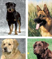
Rottweiler, German Shepherd, Golden Retriever, and Labrador Retriever (top to bottom) are breeds predisposed to coxo-femoral dysplasia. They are also the breeds most likely to be screened for this disease.
Radiographs in extension (Figure 12), as well as more specific views related to the acetabular rim (Slocum & Slocum, 1992) or joint laxity (Smith et al., 1990) can be conclusive in diagnosing joint laxity, incongruency, subchondral sclerosis and osteophyte formation.
Figure 12. Radiographs of hip joints of three different dogs.

(12A) Normal hips.
(12B) Subluxation and flattening of the femoral heads.
(12C) Severe osteophytes around the femoral heads and flattening of the acetabuli.
Epidemiology
HD is a hereditary abnormality commonly seen in certain breeds (i.e., St Bernards, Rottweilers, Newfoundlands, Bernese Mountain Dogs, German Shepherd, Labrador and Golden Retrievers) and infrequently in other breeds (i.e., Afghan Hounds, Shetland Sheepdogs, Malamutes and Huskies) (Corley, 1992).
There are 136 breeds ranked according to frequency of hip dysplasia on the Orthopedic Foundation for Animals (OFA's) website: www.offa.org. These breeds have had at least 100 evaluations between January 1974 and December 2003. The results of a retrospective study using the OFA data base have shown that there has been improvement in the hip joint phenotype of dogs in the United States. Certain breeds have shown an increase in the percentage of dogs classified as having excellent hip joint phenotype and there has been a decrease in the percentage of dogs classified as having HD. German Shepherds, Golden Retrievers, Labrador Retrievers and Rottweilers have shown the greatest increase in percentage of dogs classified as having excellent hip joint phenotype and the greatest submission screening rate. Rottweilers have shown the greatest improvement (Morgan et al., 2000). Even if these figures are biased by the tendency to pass on the best hips for official judging and not to offer the bad ones, it will help to use only the best dogs with the best hips as breeding stock.
Environmental factors, which are believed to influence the development of HD, have yet to be elucidated. Research has shown that diet has both quantitatively and qualitatively, a significant effect on the development of HD (Kealy et al., 1992). Diet will not cure HD or alter the genetic status of the offspring in this respect, but it can influence the phenotypic expression of HD by optimizing the development of the hip joints of those animals potentially at risk. Diet can also play a role in conservative treatment in those dogs where HD has already developed. Good control of body weight will alleviate clinical signs.
Pathophysiology
In the canine hip joint, both the femoral head and the acetabulum are mainly cartilaginous at birth. Bone formation, and a change in position of the femoral head in relation to the femoral shaft, will take place via endochondral ossification and osteoclastic activity respectively. In HD, joint laxity results in an incongruent hip joint where the dorsomedial part of the femoral head and the acetabular rim are in contact, while supporting almost half the body weight when walking. This causes microfractures and deformation of the acetabular rim, erosions of the cartilage and deformation of the subchondral bone (Fox et al., 1987). The associated pathological changes are joint effusion, stretching and thickening of the joint capsule and the round ligament, as well as osteophyte formation.
There are several dietary factors which play a role in hip joint development and hip joint overload, both of clinical significance in canine HD. Excess energy intake has been previously discussed. Heavy body weight will cause overloading of the cartilaginous skeleton, including the hip joints. This could be a significant factor, which may help to explain the increased frequency and severity of HD in overweight dogs.
Increased calcium intake has also been previously discussed. It can be concluded that excessive dietary calcium intake decreases maturation of the hip joint conformation as well as of the vulnerable cartilaginous template of the skeleton. This may coincide with overloading of the hip joint which is too immature in its development for the age and size of the dog and therefore, may play a significant role in deformation of the hip joint at an early age.
Hip dysplasia.
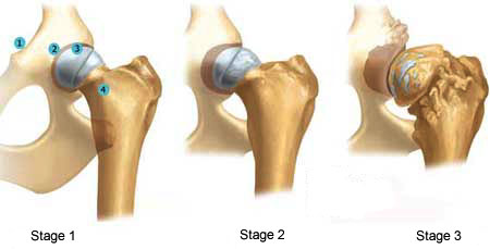
1. Pelvis
2. Femoral head
3. Femoral neck
4. Femur
In the field of canine nutrition, there is now sufficient evidence to suggest that, within the range of nutrient levels normally encountered in practice, it is not the calcium to phosphorus ratio, but the absolute calcium amount in the daily ration which determines the occurrence of skeletal abnormalities (Hazewinkel et al., 1991; Nap, 1993). A high dietary phosphorus content may bind more calcium in the intestine to form non absorbable complexes, but this is possibly only the case with non-absorbable phytates. A highly absorbable salt (as is present in bone meal) will cause the same skeletal effects as excessive calcium alone (Hazewinkel et al., 1991).
Electrolytes are present in body fluids including synovia. Differences in circulating cations (Na+, K+, Ca++ and Mg++) and anions (Cl-, H2PO4 and the SO4 present in amino acids) influence acid base balance. The influence of electrolytes on the osmolality of body fluids as well as on acid-base balance may play a role in the development of HD in young dogs.
Mean osmolality in synovial fluid from normal hip joints was significantly lower than that of synovia of hip joints of dysplastic retrievers (Olsewski et al., 1983). Whether this difference reflects the cause of joint laxity or the result of hyperperfusion of the joint capsule of the arthritic joint needs to be elucidated.
In another study (Kealy et al., 1993), the dietary content of Na+, K and Cl- ions differed (increased, increased and decreased, respectively) in the diets of three groups of dogs (n=177) of five breeds (St Bernards, German Shepherds, Coonhounds, English Pointers and Labrador Retrievers) originating from 27 litters. In these dogs, joint laxity was observed by the measurement of the Norberg angle on radiographs taken at 30 and 105 weeks of age. However, the acid-base balance and electrolyte content of body fluids were not measured. It was found that the dogs on the dry dog food (moisture <10%) with the low Na (0.32 - 0.43%), low K (0.39 - 0.70%) and high Cl (0.66 - 0.81%) content had a slight but statistically significant improvement in the Norberg angle when compared with the other groups. Only the retrievers revealed a low Norberg angle of the hip joint, irrespective of the diet. The clinical significance of these findings, the sensitivity and reproducibility of the radiographic procedure (Smith et al., 1990; Heyman et al., 1993), the influence of other electrolytes playing a role in acid-base balance and osmolality (Lemann & Lennon, 1972) must all be further investigated before the optimum electrolyte content of the food can be established. The detrimental effects of prolonged dietary-induced acidosis on skeletal mineral content (Ching et al., 1989), however, imply that further studies in this area would be valuable.
Although not proven in research, vitamin D could play a role in the development of HD. Although an increased vitamin D intake will not cause an increased absorption of calcium (see section on Vitamin D), hypervitaminosis D has a detrimental effect on the process of endochondral ossification (Tryfonidou et al., 2003b) and as such on the growth and development of the hip joint. Disturbances in cartilage differentiation may decrease the resistance of cartilage to physiological loading of the joint and lead to deformation of both femoral head and acetabular rim.
HD can develop in young, overfed dogs, even under conditions of relatively restricted activity. This is most likely due to overstressing the elasticity of the periarticular tissues and the resulting pathological cartilaginous and subchondral bone changes.
Therapy
Overnutrition should be prevented by feeding to meet the dog's energy requirements. The energy intake should be determined based on individual needs of the pet which are influenced by age, breed, body weight and activity. Since excess calcium intake can be detrimental to hip joint development, puppies should be fed diets with an appropriate calcium level for their size and age. Commercial balanced diets are available which will satisfy the special energy and calcium requirements of a fast growing puppy. These diets should never be supplemented with vitamins and minerals as excesses may occur.
Rest per se and weight loss can improve the clinical signs of HD in young and adult dogs, as was observed by force plate measurements before and after a period of 3 months cage rest (Hazewinkel, 1992).
In selected cases, development of the hip joint in young growing dogs can be surgically optimized. In dogs of 8 - 13 weeks of age, symphysiodesis is advocated by some researchers. They claim that the bottom of the pelvis does not grow in width, whereas the dorsal aspect including the acetabular roof is not hindered. As a consequence, the covering of the femoral heads will improve after thermocauterization of the symphysis pelvis. Myectomy of the pectineus muscle is indicated in dogs with contraction of these muscles, causing adduction of the hind paws even to a degree that the feet are crossing. This can be seen in young and adult dogs. Short-term results may be spectacular, whereas long term effects in regards to the development of OA are not known.
In the non surgical treatment of HD, both dietary measures and activity restriction should be employed. (© Psaila).

Other surgeries (Table 4) that can be performed when indicated include triple pelvic osteotomy (TPO), hip prosthesis and excision arthroplasty (i.e., removal of the femoral head and neck). TPO can be performed in dogs with severe hip laxity but without deformation of the head and socket. Dogs with severe hip dysplasia or severe deformation of head and/or acetabulum due to OA or trauma are potential candidates for hip prosthesis. Excision arthroplasty is indicated in cases of severe joint deformation and pain. The result of surgery is mainly dependent on the creation of smooth surface between the femur and acetabulum, the weight of the dog (<20 kg), the musculature of the dog (poorer with muscle atrophy), and early training (swimming).
Table 4. Prevention and Treatment of Different Stages of Hip Dysplasia | |
Stage of HD | Therapeutic Modalities |
Prevention | - Breed with proven HD negative parents - Prevent obesity - Prevent over-use, do not supplement a completed and balanced diet, chondroprotective agents (symphysiodesis) |
Treatment - young dogs | Adapt life style and body weight, NSAIDs, chondroprotective agents, triple pelvic osteotomy or myectomy. |
Treatment - adult dogs | Adapt life style and body weight, NSAIDs, chondroprotective agents, myectomy, hip prosthesis, excision arthroplasty. |
Orthopedic Diseases Due to Decreased Skeletal Remodeling
Decreased skeletal remodeling may occur in two separate entities: canine wobbler syndrome and enostosis, which are seen either alone or in conjunction with osteochondrosis.
Diagnosis
Ataxia, non coordinated gait of the rear legs, delayed proprioceptive reflexes and pain reaction upon extension of the neck are all signs which can be seen in young dogs of large breeds, with canine wobbler syndrome. These signs appear at the age of 6 months, unlike the unrelated ataxia in Doberman Pinchers which appears at the age of 6 years. Although not pathognomonic, the presence of the crossed extensor reflex is of great help in making the diagnosis. The other findings of a neurologic examination depend on the location of the lesion.
Shifting lameness in dogs under two years of age is suggestive of enostosis (eosinophilic panosteitis). This occurs due to the fact that all long bones are affected, but will vary in their degree of painfulness at any given time.
Plain radiographs are the initial diagnostic test. Often additional imaging techniques such as myelography and CT need to be performed to determine the exact location of the lesion in cases of canine wobbler syndrome (Figure 13). Positive pain reactions upon deep palpation of bones, together with radio-opaque areas in the medullary cavities, which arise close to the nutrient foramina, are conclusive for enostosis.
Figure 13. Disproportionate widening of the spinal canal causing compression of the spinal cord.

Epidemiology
Canine wobbler syndrome has an increased incidence in Great Danes, Mastiffs and Irish Wolfhounds and is unrelated to the spondylolisthesis and consequent ligamentous hypertrophy seen in the aged Doberman Pinscher. Enostosis occurs in a variety of dog breeds at a young age, particularly in the German Shepherd Dog.
Pathophysiology
The etiology may be multifactorial, but the influence of diet has been demonstrated in rapidly growing dogs of large breed (Hedhammar et al., 1974; Hazewinkel et al., 1985). Skeletal growth occurs in two ways: growth in length and modeling in shape. The latter includes an adaptation to changes in body size, muscle-pull and body weight. The load of hydroxyapatite crystals may cause a shift in electrons which can influence osteoblastic and osteoclastic activity. This and other still unexplained mechanisms may form the basis of Wolff's law which states that "bone is laid down where it is needed". However, the integrity of the skeleton is subordinate to calcium homeostasis, which includes the strict regulation of calcium concentration in the extracellular fluid.
As previously discussed in the section on Excess Calcium Intake, a chronic excessive calcium intake will cause high calcium absorption in young dogs, especially of large breeds (Figure 6). Calcium is not significantly excreted in the urine or via the endogenous fecal pathway, but is mainly routed to the bone in these dogs. Nutritional hypercalcitoninism induced decreased osteoclastic activity occurs with a high calcium intake and bone remodeling is diminished. As a result adaptation of the diameter of foramina to the proportional growth of the spinal cord or blood vessels may be delayed and certain forms of canine wobbler syndrome or enostosis may occur.
Great Danes fed a diet with high calcium content (i.e., 2 - 3 times the recommended amount) displayed a delayed expansion of the cervical vertebral canal in proportion to the growth of the spinal cord. Compression of the spinal cord causes myelin degeneration of both the ascending and descending tracts, the extent of which is related to the severity of clinical and imaging signs (Hedhammar et al., 1974; Hazewinkel et al., 1985).
In dogs fed high calcium diets, a decreased endosteal osteoclastic resorption, together with an increase in new periosteal bone formation has been observed (Hedhammar et al., 1974). The nutrient canals and foramina of the cortex are often abnormal in shape; this may cause edema formation, and eventually fibrosis in the medullary cavity. Edema may also extend through the cortex and underneath the periosteum, causing a loose periosteal attachment and/or excessive lamellar bone formation (Figure 14).
Figure 14. Enostosis. This 8-month-old Labrador with shifting lameness revealed pain reactions on deep palpation of the long bones including the right radius. The radiograph shows mineralization of the medullary cavity of the radius typical of enostosis and a slight thickening of the dorsal cortex of the ulna. (© HAW Hazewinkel).

Enostosis was radiologically confirmed in research animals at the age of 3 - 4 months given a gruel in their period of partial weaning (i.e., 3 - 6 weeks of age) with an elevated calcium content, together with the bitch milk. On the contrary, none of the control puppies (raised on a diet with 1% Ca DMB) first as a gruel (3 - 6 weeks) and eventually as their sole food, revealed any clinical or radiological sign of enostosis (Hazewinkel et al., 2000).
Therapy
Early dietary correction may halt the process of disproportionate remodelling of the skeleton. Commercial diets providing an adequate amount of calcium and energy for the weight and age of the dog should be instituted.
In canine wobbler syndrome, surgical decompression of the spinal cord may prevent further degeneration. Enostosis can be very painful and recurrent. Non-steroid anti-inflammatory drugs (NSAIDs) can be prescribed. Enostosis will heal without long term effects however, relapses may occur until the dog is 2 years of age.
About
How to reference this publication (Harvard system)?
Affiliation of the authors at the time of publication
1Utrecht University, Utrecht, Netherlands.2South Pasadena, CA, USA.

Comments (0)
Ask the author
0 comments