
Fringe-eared Oryx (Oryx gazella callotis)
Read
Order: Artiodactyla
Family: Bovidae
1) General Zoological Data
There are three oryx species (Oryx dammah, Oryx gazella, and Oryx leucoryx). Of the South African Oryx gazella, Grubb (1993) recognized 11 or 12 subspecies, which includes the animal under discussion. The fringe-eared oryx locates primarily to East Africa, Kenya and adjacent regions. A substantial colony has been bred at the San Diego Zoo.
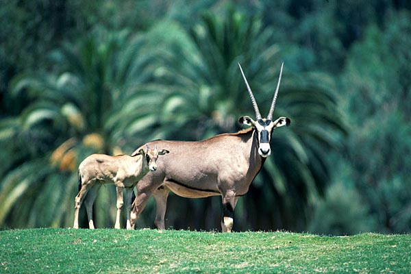
Fringe-eared oryx at San Diego Wild Animal Park.

Fringe-eared oryx and offspring.
2) General Gestational Data
Neonates weigh around 9-11 kg; first births are expected at two years of age. Gestation lasts about 265 days and singletons are the norm.
3) Implantation
No early implantational stages have been observed and this new gestation is too young to qualify.
4) General Characterization of the Placenta
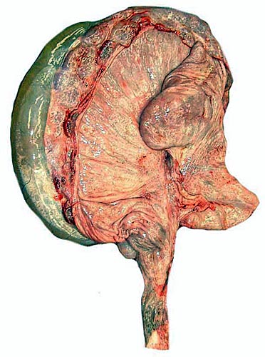
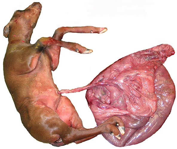
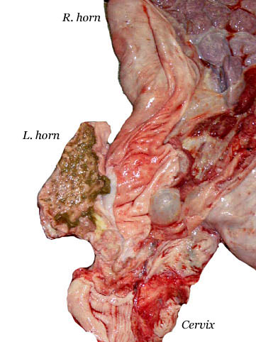
I have had the opportunity of studying the uterus, fetus and placenta of three pregnant animals. One dam died from peritonitis secondary to a penetrating wound. The gestation was near term. The opened uterus, with fetus exposed, is shown first. The second dam was euthanized because of a broken foot and osteomyelitis. She was pregnant with a less advanced female fetus.
The male fetus of the first gestation weighed 4,625 kg (considerably less than a full term neonate is expected to weigh) and it was located in the right uterine horn. Its CR length was 48 cm, and it was normally developed. The left horn was empty and the placenta did not extend into it. That uterine horn was considerably smaller. There were 100 cotyledons of a moderately convex type. They varied from 4 to 9 cm in diameters. After detaching the placenta from the uterus, it weighed 1,200 g.
The second gestation was less advanced but better preserved. It had a fetus weighing 2,250 g with a crown-rump length of 38 cm. The location was the right uterine horn again, and the placenta did not extend into the second horn. The corpus luteum was also on the right. This placenta had 120 cotyledons that varied in size between 2 and 8 cm. The whole specimen was 50 x 50 cm and, including the attached uterus, the specimen without fetus weighed 2.335 kg. The dam weighed (pregnant) 120 kg.
The third gestation was considerably younger and the dam died post anesthesia with pulmonary edema. The gestation occurred again in the right uterine horn; the male fetus was 10 cm CR length and weighed 51.6 g. Only 55 cotyledons were found in this placenta but many were so small (<1 cm) that there may well have been more.
First fringe-eared oryx fetus and placenta. Note the short, straight umbilical cord and its numerous yellow granules that extend onto the amnion.

Uterus of oryx partially opened and exposing a large allantoic sac (left), a few cotyledons and (on top right) the smaller empty uterine horn.
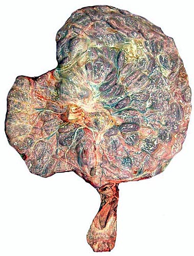
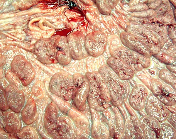
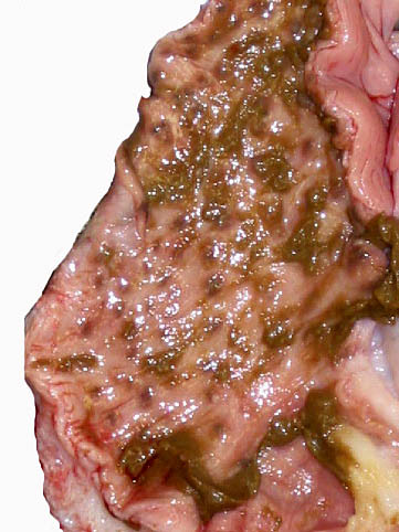
Uterus fully opened and fetus removed showing the insertion of the umbilical cord and the large number of closely packed cotyledons.
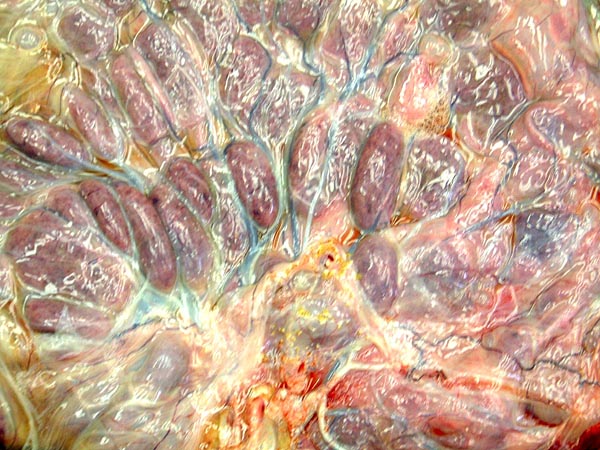
Close-up of cord insertion site showing the yellow areas of squamous metaplasia and a row of slightly convex cotyledons.
Uterus after removal of the placenta with somewhat irregularly arranged caruncles.

Close-up of uterine caruncles.
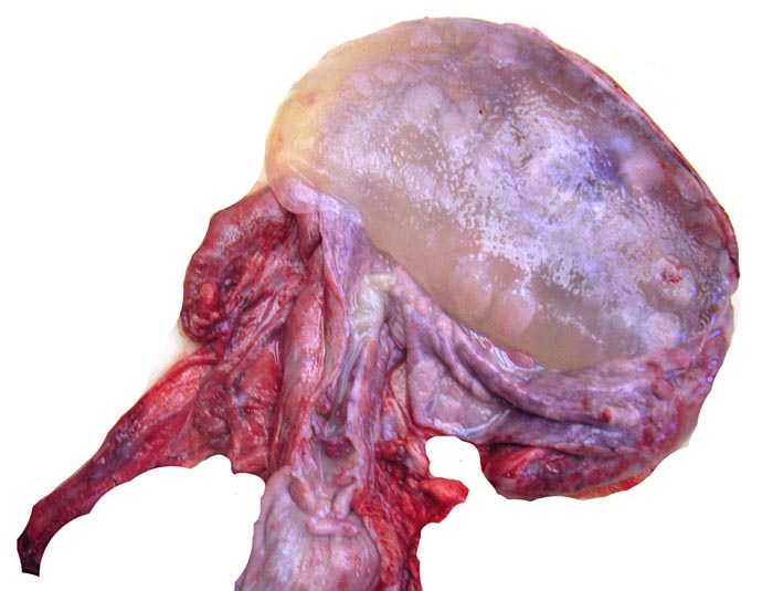
Second pregnant oryx uterus with ovaries at white arrows and fetus located in right horn. The long extension below is the cervix and portion of vagina.

Opened uterus of second gestation with large allantoic sac intact at left.

Opened right horn of the second pregnant uterus with large number (120) of cotyledons arranged in four parallel rows.

Third gestation with pregnancy in right horn.

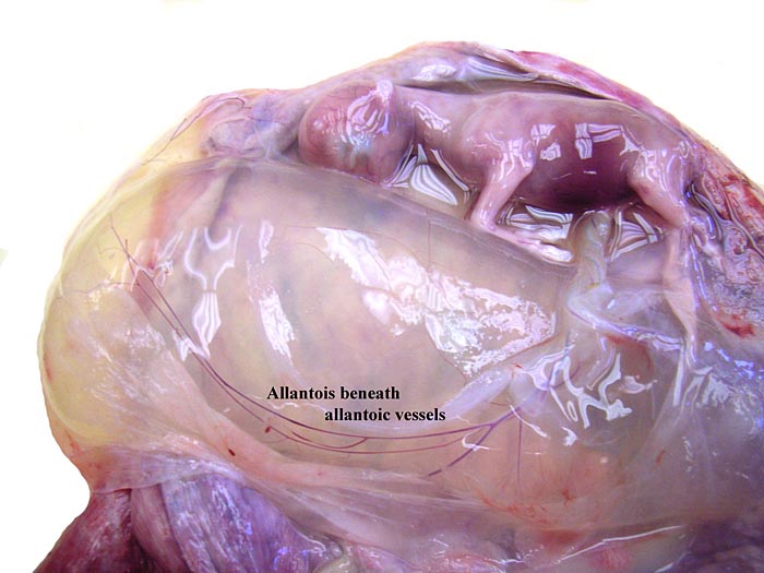
Right horn opened showing the allantoic sac.
Same gestation with amnion opened and allantoic sac with its vessels exposed.
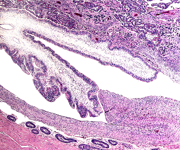
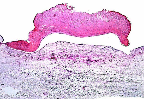
Implanted fringe-eared oryx placenta with uterine wall below.

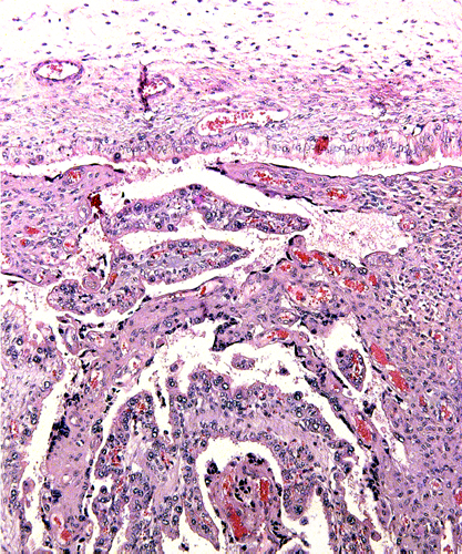
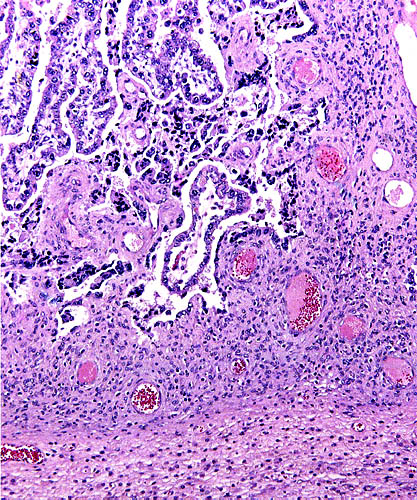
Edge of placenta with columnar trophoblast of allantochorion apposed to the relatively inactive endometrium below.
Beneath the chorionic membranes is cylindrical trophoblast with relatively sparse pigmentation.

Placental surface of second, better-preserved placenta which had no pigment in the trophoblast.
5) Details of Fetal/maternal Barrier
This typically cotyledonary placenta is epithelio-chorial. A major dispute is the origin of the trophoblastic pigment beneath the chorionic surface. Much further discussion is had in the chapters on deer and sheep. Suffice it to say here, that it is NOT iron-containing pigment, nor is it bilirubin, but it appears to be melanin or a related compound. Binucleated trophoblastic cells are present. None of this pigment was observed in the third, young, gestational sac.
High magnification of the epithelio-chorial fetal/maternal "barrier" with a thin capillary running in loose fetal villous connective tissue above.

6) Umbilical Cord
The umbilical cords measured 12 cm and 19 x 2.5 cm respectively, had no spirals and were covered with numerous small yellow granules of squamous metaplasia. They contained four large blood vessels and a central allantoic duct. In another fetus shown next, the amnionic portion of umbilical cord measured 16 cm in length and had a slight right spiral. The third fetus had a 3 cm long unspiraled cord with large allantoic duct. The latter was surrounded by tiny vessels and the surface had already numerous squamous plaques.
Stillborn fringe-eared oryx placenta and uterus from dead dam.
Umbilical cord with two arteries (left) and veins (right). Collapsed allantoic duct in center. Note the squamous callosities on the surface at right.
Insertion of the umbilical cord (second uterus) with yellow granules of squamous metaplasia. The filled allantoic sac can be seen beneath.

One of the numerous yellow surface regions of focal keratinization of the cord's amnionic surface epithelium.
The allantoic duct in the center of the umbilical cord has some musculature in its wall and numerous small blood vessels.

7) Uteroplacental Circulation
The nature of uterine circulation is as yet unknown.
8) Extraplacental Membranes
The allantoic sac of these gestation were huge and filled with yellow urine. They also contained a small amount of hippomanes. The amnion was closely applied to the fetus. It had the same yellowish granules, especially near the insertion of the umbilical cord, as can bee seen from the first placental photograph. They are areas of squamous metaplasia. Remnants of vitelline structures were not found.
Thin membranes to show focus of squamous metaplasia, extensive wrinkling of amnionic surface, and very thin allantoic epithelium in this markedly distended allantoic sac.

9) Trophoblast External to Barrier
At the site of implantation the trophoblast infiltrates the superficial endometrium, surrounds the maternal blood vessels, but does not invade them. The trophoblast does not penetrate into the myometrium either.
Placental floor of the second placenta. Endometrium (without glands) is at the base and is superficially infiltrated by the more purple trophoblast that surrounds the maternal blood vessels.
10) Endometrium
There is no decidual formation. The endometrium of pregnancy assumes a relatively fibrous appearance. The changes in the unoccupied horn are quite remarkable. It is separated from the pregnant horn and is very much smaller. It contains numerous small caruncles, all of which are green and soft. The second pregnancy had a remarkable amount of pasty green, degenerated material in this second horn. The material stains intensely blue but contains no crystals. The endocervical canal is filled with a thick mucous plug that interdigitates with the endocervial glands (see last picture). The importance of this mucus cannot possibly be undervalued as it prevents ascending infection.
Second pregnancy with opened left uterine horn.
Empty left uterine horn of the first uterus showing the discolored (greenish) small caruncles.

Empty left horn of second gestation. Much soft, green material was contained in this endometrial cavity.
This is the unused horn of the second pregnant with much green (here blue) degenerated debris on the surface of inactive endometrium.

Representative caruncle from the empty uterine horn. Note the extensive vasculature.

11) Various Features
No subplacenta exists but the endometrium of the caruncles show an extensive vascular development.
12) Endocrinology
Patton et al. (2001) studied the control of aggression in a bachelor herd of fringe-eared oryx by administering melengestrol. Fecal androgen excretion decreased significantly, as did aggression. The corpora lutea were all in the right ovary which was suitably enlarged; in the third pregnancy the ovary measured 4 cm in greatest diameter, while the left was 2 cm.
13) Genetics
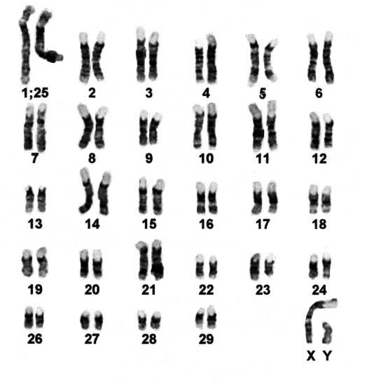
The fringe-eared oryx has 58 chromosomes (Kumamoto et al., 1999). They are very similar in karyotype to those of the other forms of the genus Oryx. A hybrid of fringe-eared oryx and a beisa oryx (Oryx gazella beisa) was reported by Gray (1972).
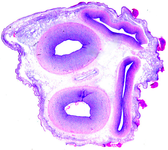
Giemsa-banded chromosomes of Fringe-eared oryx (2n=58).
14) Immunology
No studies are known to me. It is important, however, to point out that most animals, including this oryx, produce enormous quantities of a very thick, tenacious mucus in the endocervical canal during pregnancy. This is essential as it protects against ascending infection from the vagina. The mucus contains many cytokines, antibodies and is also a physical barrier (see Hein et al., 2001 for a study of human cervical mucus).
This is a section of the endocervix from the second specimen showing intense mucus production.

15) Pathological Features
Griner (1983) had extensive experience with oryx species and reported most deaths as being due to trauma, some cases of "bloat" and hemangiomas.
16) Physiologic Data
There have been no studies.
17) Other Resources
Numerous cell strains of this and related oryx species are available from CRES through contacting Dr. O. Ryder at [email protected].
18) Other Remarks - What Additional Information Is Needed?
Virtually no endocrinology has been done in this species and the weight and appearance of term placentas are unknown. Early implantational stages should be studied.
Acknowledgement
The animal photographs in this chapter come from the Zoological Society of San Diego. I appreciate also very much the help of the pathologists at the San Diego Zoo.
Gray, A.P.: Mammalian Hybrids. A Check-list with Bibliography. 2nd edition. Commonwealth Agricultural Bureaux Farnham Royal, Slough, England, 1972.
Griner, L.A.: Pathology of Zoo Animals. Zoological Society of San Diego, San Diego, California, 1983.
Grubb, P.: Order Artiodactyla, pp. 377-414, in Mammal Species of the World, 2nd ed., D.E. Wilson and D.A.M. Reeder, eds. Smithsonian Institution Press, Washington, 1993.
...About
How to reference this publication (Harvard system)?
Affiliation of the authors at the time of publication
Department of Reproductive Medicine and Pathology, School of Medecine, University of California, San Diego, CA, USA.

Comments (0)
Ask the author
0 comments