
Slenderhorned Gazelle (Gazella leptoceros leptoceros)
Read
Order: Artiodactyla
Family: Bovidae
1) General Zoological Data
Slender-horned gazelles ("Dunengazellen" in German, also known as "Rhime" and Loder's gazelles) are now rare, endangered gazelles that once ranged in the desert from Algeria to Egypt (Saleh, 1987). Although paler and slightly larger, this species can be mistaken for Dorcas gazelles (Newby, 1984; see also my chapter on Dorcas gazelles). Groves (1969) found greater similarity to the goitered gazelle in his meticulous comparative skeletal study, and hybridization between the two forms has been described. Distinguishing features are the horns. The slender-horned gazelles are very small animals, weighing 14-18 kg and they have been maintained in only a few zoos. Those animals come from a very small founder stock of individuals (Thomas et al., 1986). For this reason, inbreeding is a problem for the captive population. At the San Diego, as in some other locations, numerous births have occurred. The longevity of slender-horned gazelles is about 11 years, and age of first parturition is at about 11-12 months (Mentis, 1972).
The evolutionary history of Bovidae and of also that of many gazelles is still controversial. It was addressed with modern DNA studies by Gatesy et al. (1997), and also by Matthee & Davis (2001). The chromosomal study by Effron et al. (1976) suggested that, because of the autosome/X fusion of this and many other African gazelles, they have a common ancestor, despite their current great variability in phenotypes and karyotypes.
There have been suggestions in the scientific literature that one should distinguish two, perhaps three subspecies of slender-horned gazelles: G. l. leptoceros, G. l. loderi, and perhaps G. marica. Detailed studies of these putative relationships may be found in the study conducted by Lange (1972).
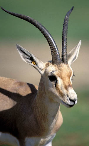
Slender-horned gazelle at San Diego's Wild Animal Park.
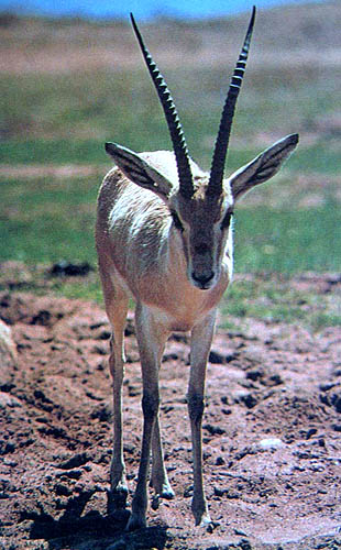
Male and female slender-horned gazelles.

Gazella leptoceros.
2) General Gestational Data
Slender-horned gazelles have a gestational length of around 165-169 days (Dittrich, 1968). Newborns weigh between 1 and 1.8 kg. The adult female of the specimen shown here, weighed 12.5 kg and died because of trauma that was sustained during a fight occurring during her gestation. The male fetus was 10 cm long and weighed 28 g. The entire specimen shown here, uterus and vagina included, weighed 105 g. There is generally only one young born; however, at San Diego's Wild Animal Park, twins were born and raised in 1971, of some 30 animals that had been produced by then.
3) Implantation
The implantation of this immature placenta was mesometrial. The nonpregnant horn had approximately 40 caruncles in four rows and, in the specimen available, it was filled with tenacious mucus. The corpus luteum was on the side (left) of the gestation. The right ovary was significantly smaller and had no significant luteinization. The time of implantation has yet to be defined for this species.
4) General Characterization of the Placenta
Slender-horned gazelles have a bicornuate uterus and their placenta is epithelio-chorial, villous and polycotyledonary. The villi are branched; the trophoblast is similar to that of most other bovidae. They apparently implant mostly in the left uterine horn, as witnessed also by the absence of corpora atretica in the right ovary.
Uterus and vagina of pregnant slenderhorned gazelle. The fetus and placenta are in the left uterine horn.
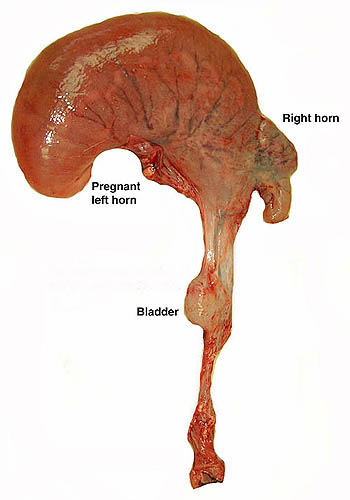
The same specimen viewed from anterior.

Opened uterus with female fetus (10 cm) in left horn (here seen at the right). Note the white, granular seams of the concave cotyledons. Endometrial caruncles are seen in the right horn.

The same gestation with the fetus moved to expose the cotyledons.
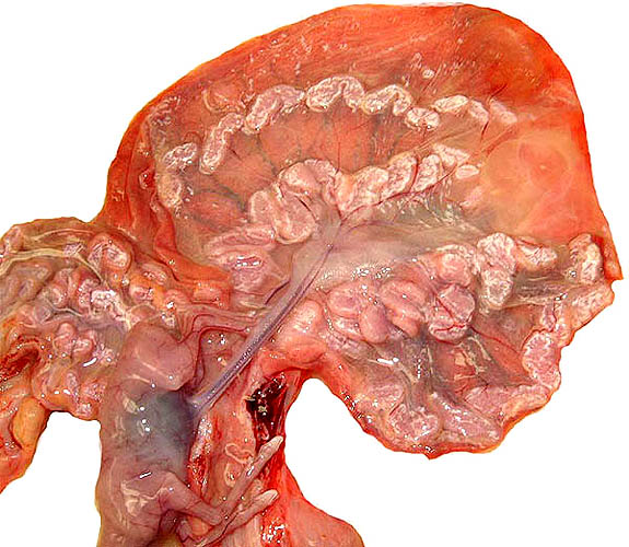
Two gestations were available to me for study. One was a uterus with male fetus of 10 cm length in its placenta, and attached to the uterus. There were 40 cotyledons in four rows, as shown above. Some of these cotyledons were fused and were thus difficult to enumerate precisely. The cotyledons had a central dimple (concavity) and a peripheral seam of white granular material. This white granular material did not refract with polarizing filters and is assumed to represent the cellular debris at the edge of all cotyledons, at the place where the intercotyledonary membrane is folded over areolae (see histology below). The placenta is chorioepithelial in nature and not invasive.
The second placenta came from a term gestation. It weighed 70 g and had a 10 cm umbilical cord attached. It possessed 57 ellipsoid cotyledons that measured between 0.5 and 2.5 cm. The greatest dimensions of this specimen were 17 x 4 cm. As is true of all delivered placentas, the maternal intercotyledonary endometrial tissue was absent and the organ was slightly autolyzed.
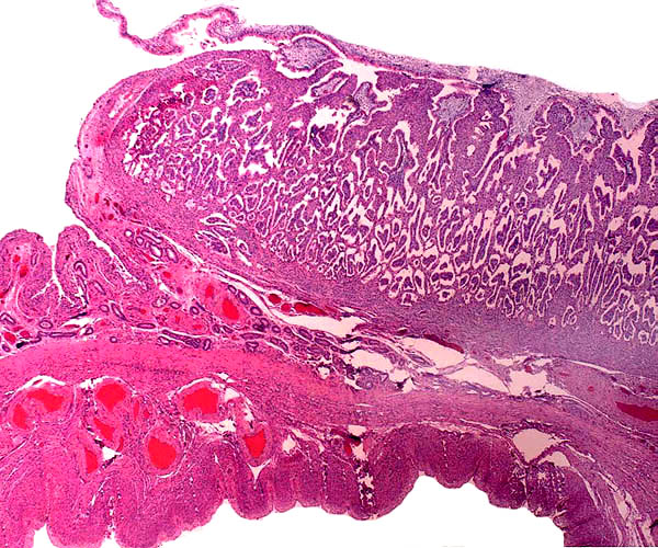
Edge of immature cotyledon attached to myometrium below. The intercotyledonary endometrium is folded and glandular. The ingrowths of villi (purple-blue centers) from chorioallantoic membrane are obvious.
5) Details of fetal/maternal barrier
During the process of implantation, the crevices of the caruncles (to be shown subsequently in the nonpregnant horn) are being infiltrated by the chorioallantoic membrane connective tissue's villous extensions and their trophoblastic covers. Subsequently, the villi come to interdigitate diffusely with the caruncles.
This is the surface of the immature placenta with villi (having small fetal capillaries) interdigitating with reddish endometrium.

The "floor" of the implantation of the cotyledon shows few endometrial glands and a markedly thickened, fibrous endometrium. The fetal villi are the structures within the spaces. There is no trophoblastic infiltration of the endometrium.

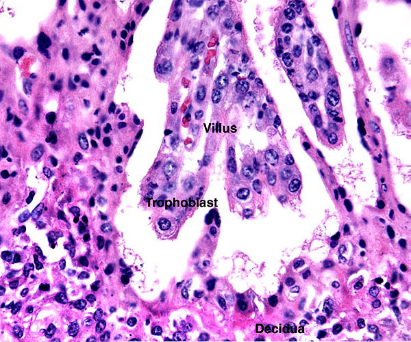
Two immature villi interdigitate with endometrium (below). Note the giant nuclei of the endometrial epithelium.
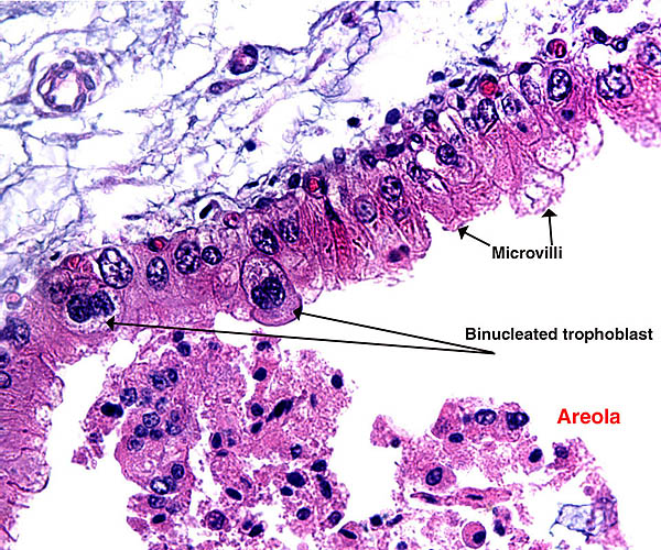
The trophoblast that covers the villi of the slender-horned gazelle placenta is cuboidal to columnar and has numerous microvillous projections. Because of shrinkage artifacts, it is pulled away from the intervening endometrium in the microphotographs shown, leaving a space that has some proteinaceous material within it. Many trophoblast nuclei are somewhat hyperchromatic and they are perhaps hyperdiploid, but true giant cells do not occur. There are also typical trophoblastic binucleate cells, which are so characteristic of so many ruminants and which are believed to produce relaxin. They are most numerous and best developed in the tall columnar trophoblast of the membranes that stretches between the caruncles. They overly much debris and protein. Some of this is presumably due to the secretory activity of the endometrial glands in those locations. This secretion is referred to as "uterine milk". Amoroso (1961) has presented some data on the composition of uterine milk in cow, sheep and mare. Around 10% is protein, 1.5% is lipids.
The endometrium to which the villi are loosely apposed is somewhat fibrous in appearance and has a complex mixture of cells. On the surface that is exposed to trophoblast, the endometrial epithelial cells are flattened and often have hyperchromatic, enlarged nuclei.
The fetal capillaries of the slightly branched villi are not numerous. They lie predominantly beneath the trophoblast, without indenting it.
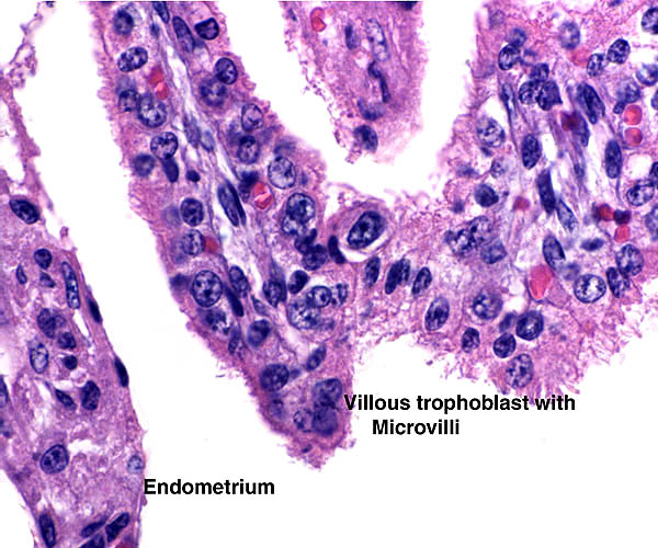
Tip of immature villus with microvillous trophoblastic surface and some giant nuclei.
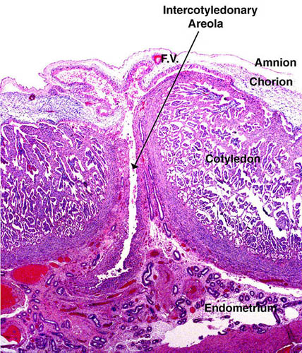
Two adjacent immature cotyledons are separated by a cleft that corresponds to an "areola", i.e. endometrial tissue with secretory activity. (F.V.=Fetal Vessel).
Trophoblast under the chorioallantoic membrane that overlies an areola. This shows strikingly the many binucleated trophoblastic cells. The debris and secretion in the areolar cavity is obvious (below). This is probably responsible for the granular, white deposits at the edge of the cotyledons.
Term, delivered placenta of a slender-horned gazelle. This specimen, of course, has no maternal septa, and consists of villi and chorion (above) only.
6) Umbilical cord
In the immature specimen described here, the umbilical cord was 5 cm long and had no twists. It contained four large blood vessels and an allantoic duct. The connective tissue adjacent to the allantoic duct had several small blood vessels, while none were found in the other regions of the umbilical cord. There were a few caruncles composed of squamous metaplasia. The term umbilical cord was 10 cm in length, also had four vessels and a small allantoic duct, but there were more numerous small and more muscular additional vessels in the cord that were not confined to the allantoic duct. The duct had taller epithelium than found in the immature specimen.
Umbilical cord of immature specimen. Two arteries and two veins, allantoic duct (A.D.) and foci of squamous metaplasia on the surface.

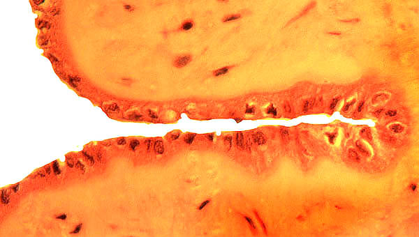
Allantoic duct of immature specimen with tiny allantoic blood vessels in the fibromuscular wall of the allantoic duct.

The allantoic duct epithelium of a term umbilical cord is taller.
7) Uteroplacental circulation
No such studies have been conducted.
8) Extraplacental membranes
The amnionic membranes were filled with clear fluid (urine) in the immature specimen. The amnionic epithelium was flat to minimally cuboidal. There are no blood vessels. The amnion has numerous small caruncles which are composed of squamous metaplasia. Vitelline tissue was absent from both specimens. Also, no allantoic sac was identified in the implanted specimen, nor were there membranes histologically resembling an allantoic sac. No hippomanes were present.
Focus of squamous metaplasia on amnion of the immature specimen.

9) Trophoblast external to barrier
There is no invasion of the endometrium by the trophoblast. Although some trophoblast has large, hyperchromatic nuclei, no true giant cells are present.
10) Endometrium
The endometrium of pregnancy differs in the different regions of the uterus. Normal glandular tissue is present between the caruncles but, beneath the caruncles, the endometrial stroma is more fibrous and has very few glands. No true decidual cells, such as seen in primate placentas, are found. In the endometrium beneath the cotelydonary implantations, the surface epithelial cells of the endometrium are flat and have hyperchromatic nuclei. The caruncles are not shed at delivery of the placenta; the villi are merely being peeled out of the crypts.
The non-pregnant right horn of this immature uterus had caruncles such as seen here. The cavitary nature of the caruncle is apparent. Placental villi would grow into these crevices as was shown earlier.
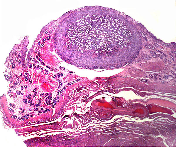
Higher magnification of a (non-pregnant) caruncle.

11) Various features
No other remarkable features have been observed. A subplacenta does not exist.
12) Endocrinology
The fetal testis had numerous stimulated interstitial cells.

The fetal testis shown above exhibits a moderate stimulation of its interstitial cell component. This suggests that some LH-like activity occurs in utero, but it is not necessarily derived from the placenta. I have found no endocrine studies on slender-horned gazelles in the literature; however, some information has been gathered on the related goitered gazelle (Sempere et al., 2001). Prolactin and progesterone determinations were carried out in two different environments by these investigators.
13) Genetics
Slender-horned gazelles are among those gazelles with a translocation of the X-chromosome to an autosome (Effron et al, 1976). Thus, males have 33 chromosomes, females have only 32. The amount of DNA, however, is the same in the two sexes. Hybrids between G. leptoceros marica and G. subgutturosa have been described in London (Gray, 1972).
Genetic studies on slender-horned gazelles are few in number, while other species have had closer scrutiny. Thus, electrophoretic and chromosomal studies were done on three related species and then compared with findings from G. subgutturosa (Vassart et al., 1993 & 1995a, b).
14) Immunology
I have found no immunological studies on slender-horned gazelles.
15) Pathological features
Griner (1983) listed as the commonest cause of death the sequelae of trauma and fights. He also depicted a fatal case of avian tuberculosis. In addition, Griner described a neonatal death that was due to severe icterus whose etiology remained undetermined. Intestinal parasites were reported by Stover & Dolensek (1985). Stover et al. (1990) found toxoplasmosis in a slender-horned gazelle at autopsy; another animal had antibody titers but survived. Nocardia and Actinomyces were found in six gazelles. This apparent susceptibility was attributed to possible inbreeding effects (Kinde et al., 1992).
16) Physiologic data
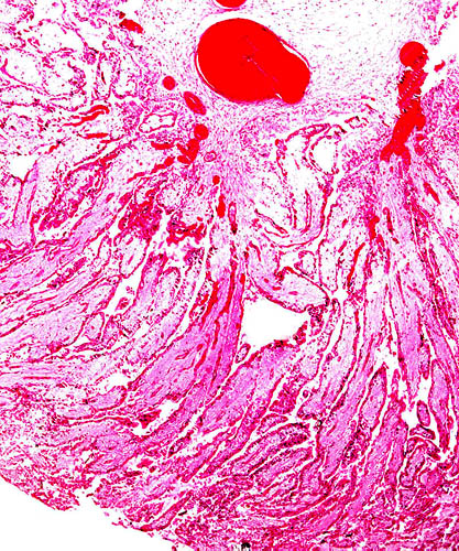
The presence of much mucus within the nonpregnant uterine horn and in the endocervical canal (shown below) is significant. The endocervical mucus surely serves to occlude the cervical os during gestation and it thus prevents ascent of organisms.
Vaginal-cervical junction. Endocervix at right, vagina at left. Note the intense mucus production of endocervical glands in pregnancy.

Stover & Dolensek (1985) reported the successful blood transfusions from a Dorcas gazelle to a 6.8 kg, two-months-old slender-horned gazelle that was sick and anemic from intestinal parasites (Haemonchus sp.). General laboratory data on this species were published by ISIS (1997).
17) Other resources
Cell lines are available from CRES at San Diego Zoo by contacting Dr. Oliver Ryder at: oryder@ucsd.edu.
18) Other remarks - What additional Information is needed?
Only one mature placenta has been studied, and the length of only one term umbilical cord is known. More specimens need be collected, and endocrinological studies are needed.
Acknowledgement
The animal photographs in this chapter come from the Zoological Society of San Diego. I appreciate also very much the help of the pathologists at the San Diego Zoo.
Amoroso, E.C.: Placentation. Chapter 15, pp.127-311, In, Marshall's Physiology of Reproduction, V.II, A.S. Parkes, ed. Second Edition. Little, Brown & Co. Boston, 1961.
Dittrich, L.: Keeping and breeding gazelles at Hannover Zoo. Intern. Zoo Ybk. 8:139-143, 1968.
Effron, M., Bogart, M.H., Kumamoto, A.T. and Benirschke, K.: Chromosome studies in the mammalian subfamily Antilopinae. Genetica 46:419-444, 1976.
...About
How to reference this publication (Harvard system)?
Affiliation of the authors at the time of publication
Department of Reproductive Medicine and Pathology, School of Medecine, University of California, San Diego, CA, USA.

Comments (0)
Ask the author
0 comments