menu sluit menu
Home Home
Login
Main navigation
  • Library
  • Calendar
  • e-Learning
  • News
    • Veterinary News In this section you find veterinary news
    • Recent Additions All content that was recently added to the IVIS library
  • Get involved
    • Donate Support IVIS, make a donation today
    • Media kit Promote your e-learning & events on IVIS
    • Add your e-learning & events to the IVIS calendar
    • Publish on IVIS Publish your work with us
  • About
    • Mission Our Mission Statement
    • What we do More info about IVIS and what we do
    • Who we are More info about the IVIS team
    • Authors See list of all IVIS authors and editors
  • Contact
User tools menu
User tools menu
Main navigation
  • Library
  • Calendar
  • e-Learning
  • News
    • Veterinary News In this section you find veterinary news
    • Recent Additions All content that was recently added to the IVIS library
  • Get involved
    • Donate Support IVIS, make a donation today
    • Media kit Promote your e-learning & events on IVIS
    • Add your e-learning & events to the IVIS calendar
    • Publish on IVIS Publish your work with us
  • About
    • Mission Our Mission Statement
    • What we do More info about IVIS and what we do
    • Who we are More info about the IVIS team
    • Authors See list of all IVIS authors and editors
  • Contact
Follow IVIS
  • Twitter
  • Facebook
Support IVIS

Breadcrumb

  1. Home
  2. Library
  3. European Veterinary Conference - Voorjaarsdagen
  4. EVC - Voorjaarsdagen - The Hague, 2017
  5. Performing and interpreting sinus endoscopy
European Veterinary Conference - Voorjaarsdagen
Back to Table of Contents
Add to My Library
Close
Would you like to add this to your library?

Get access to all handy features included in the IVIS website

  • Get unlimited access to books, proceedings and journals.
  • Get access to a global catalogue of meetings, on-site and online courses, webinars and educational videos.
  • Bookmark your favorite articles in My Library for future reading.
  • Save future meetings and courses in My Calendar and My e-Learning.
  • Ask authors questions and read what others have to say.
Sign in Register
Comments
Share:
  • Facebook
  • LinkedIn
  • Mail
  • Twitter

Performing and interpreting sinus endoscopy

Author(s):
Pascoe R.J.
In: EVC - Voorjaarsdagen - The Hague, 2017 by European Veterinary Conference - Voorjaarsdagen
Updated:
JUL 05, 2017
Languages:
  • EN
Back to Table of Contents
Add to My Library
Close
Would you like to add this to your library?

Get access to all handy features included in the IVIS website

  • Get unlimited access to books, proceedings and journals.
  • Get access to a global catalogue of meetings, on-site and online courses, webinars and educational videos.
  • Bookmark your favorite articles in My Library for future reading.
  • Save future meetings and courses in My Calendar and My e-Learning.
  • Ask authors questions and read what others have to say.
Sign in Register
SHARE:
  • Facebook
  • LinkedIn
  • Mail
  • Twitter
    Read

    Equine sinus disease is a common condition of the equine head. Typically it presents with a unilateral nasal discharge. The characteristics of the discharge may give clues to the nature of the disease. Nasal endoscopy should be the first step to identify the origin of any nasal discharge. The drainage angle of the rostral and caudal maxillary sinuses lie rostral to the ethmoturbinates in the middle meatus. Whilst it is possible to perform transnasal sinus endoscopy the size of the drainage angle means smaller scope diameters are required. In cases with sinusitis the drainage angle may be enlarged making endoscopy easier, however, in many cases access will not be possible.

    Radiography can be useful to assess the sinuses for presence of fluid or soft tissue opacity but it can be difficult to determine the exact location and nature of these. Standing computed tomography has more recently been used to assess conditions of the head. The three-dimensional nature of this imaging modality removes problems associated with superimposition and can increase the sensitivity and specificity of diagnostic imaging. However, the availability of the modality is comparatively limited and can be expensive. Systems capable of computed tomography in the anaesthetized horse may be more widely available but there is an increased risk and cost. Undoubtedly computed tomography offers the best opportunity to recognize disease and anatomical variation in the individual case.

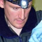
    Direct sinus endoscopy via trephination offers the opportunity to visualize the internal structure and contents of the sinus cavities. The sinuses are divided in to seven paired compartments which are separated in to two groups, rostrally and caudally. The rostral group comprises the rostral and ventroconchal sinuses. The caudal group is more complicated comprising frontal, dorsal conchal, caudal maxillary, sphenopalatine and ethmoidal sinuses. The frontal and dorsal conchal are sometimes referred to as the conchofrontal sinus.

    Rostral maxillary sinus portal

    The rostral maxillary sinus can be trephined directly 1cm below the halfway point on a line between the medial canthus and infraorbital foramen. Access is limited, especially in younger horses, due to the apices of the cheek teeth and therefore it is rarely used. The ventroconchal sinus cannot be accessed from this portal. It may be useful for specific conditions, such as cementoma, particularly following computed tomography to identify a specific site to allow treatment.

    Caudal maxillary sinus portal

    The caudal maxillary sinus can be trephined 2cm ventral and rostral to the medial canthus of the eye. This provides access to caudal sinus group. From here the maxillary septal bulla can idenitified.

    Conchofrontal sinus portal

    The conchofrontal sinus can be trephined caudal to a line between the medial canthi, slightly lateral to a midpoint between the ipsilateral medial canthus and midline of the skull. This provides access to the caudal sinus group including frontal and dorsal conchal sinuses and via the frontomaxillary opening the caudal maxillary sinus, sphenopalatine sinus and the maxillary septal bulla.

    Maxillary septal bulla fenestration

    Access from the caudal sinus group in to the rostral group can be achieved by fenestrating the maxillary septal bulla. This overlies the infraorbital canal and is a thin dividing wall between the two. In most cases this is most easily achieved via the conchofrontal sinus portal which then affords access in to the rostral maxillary and ventroconchal sinuses. In around 10% of cases the bulla cannot be fenestrated via this approach and access has to be created by a caudal maxillary sinus portal.

    [...]

    https://voorjaarsdagen.eu/file/2019/07/Rob-Pascoe-EQ-PERFORMING-AND-INTERPRETING-SINUS-ENDOSCOPY.pdf
    Back to Table of Contents
    Add to My Library
    Close
    Would you like to add this to your library?

    Get access to all handy features included in the IVIS website

    • Get unlimited access to books, proceedings and journals.
    • Get access to a global catalogue of meetings, on-site and online courses, webinars and educational videos.
    • Bookmark your favorite articles in My Library for future reading.
    • Save future meetings and courses in My Calendar and My e-Learning.
    • Ask authors questions and read what others have to say.
    Sign in Register
    Comments (0)

    Ask the author

    0 comments
    Submit
    Close
    Would to like to further discuss this item?

    Get access to all handy features included in the IVIS website

    • Get unlimited access to books, proceedings and journals.
    • Get access to a global catalogue of meetings, on-site and online courses, webinars and educational videos.
    • Bookmark your favorite articles in My Library for future reading.
    • Save future meetings and courses in My Calendar and My e-Learning.
    • Ask authors questions and read what others have to say.
    Sign in Register
    About

    How to reference this publication (Harvard system)?

    Pascoe, R. (2021) “Performing and interpreting sinus endoscopy”, EVC - Voorjaarsdagen - The Hague, 2017. Available at: https://www.ivis.org/library/evc/evc-voorjaarsdagen-hague-2017/performing-and-interpreting-sinus-endoscopy (Accessed: 24 March 2023).

    Author(s)

    • Rob Pascoe

      Pascoe R.J.

      BVSc BAEDT DiplAVDCEq MRCVS
      Bell Equine Veterinary Clinic,
      Read more about this author

    Copyright Statement

    © All text and images in this publication are copyright protected and cannot be reproduced or copied in any way.
    Related Content

    Readers also viewed these publications

    • Proceeding

      BEVA - Annual Congress - Liverpool, 2022

      By: British Equine Veterinary Association
      MAR 20, 2023
    • Journal Issue

      Veterinary Evidence - Vol 8 N°1, Jan-Mar 2023

      In: Veterinary Evidence
      MAR 19, 2023
    • Proceeding

      AVEF - Conférence Annuelle - Reims, 2022

      By: Association des Vétérinaires Équins Français
      MAR 03, 2023
    • Proceeding

      EEHNC - Virtual Congress - 2021

      By: European Equine Health and Nutrition Congress
      FEB 09, 2023
    • Proceeding

      SFT - Theriogenology Annual Conference - Bellevue, 2022

      By: Society for Theriogenology
      JAN 10, 2023
    • Proceeding

      ACVIM & ECEIM - Consensus Statements

      By: American College of Veterinary Internal Medicine
      NOV 11, 2022
    • Journal Issue

      Veterinary Evidence - Vol 7 N°2, Apr-Jun 2022

      In: Veterinary Evidence
      OCT 07, 2022
    • Journal Issue

      Veterinary Evidence - Vol 7 N°3, Jul-Sep 2022

      In: Veterinary Evidence
      OCT 04, 2022
    • Journal Issue

      Veterinary Practice Management Articles - Veterinary Focus

      In: Veterinary Focus
      AUG 05, 2022
    • Chapter

      Nutrition

      In: The Clinical Companion of the Donkey (2nd Edition)
      JUL 09, 2022
    • Chapter

      Lymphatics and Lymph Nodes

      In: Current Techniques in Small Animal Surgery (5th Edition)
      JUL 08, 2022
    • Chapter

      Pharmacology and Therapeutics

      In: The Clinical Companion of the Donkey (2nd Edition)
      JUL 03, 2022
    • Chapter

      Heart and Great Vessels

      In: Current Techniques in Small Animal Surgery (5th Edition)
      JUN 27, 2022
    • Chapter

      Sedation, Anaesthesia and Analgesia

      In: The Clinical Companion of the Donkey (2nd Edition)
      JUN 05, 2022
    • Chapter

      The Geriatric Donkey

      In: The Clinical Companion of the Donkey (2nd Edition)
      MAY 20, 2022
    • Chapter

      Euthanasia and the Post-Mortem Examination

      In: The Clinical Companion of the Donkey (2nd Edition)
      MAY 20, 2022
    • Chapter

      Endocrine System

      In: Current Techniques in Small Animal Surgery (5th Edition)
      MAY 15, 2022
    • Chapter

      Appendix 7: Example Diets: for the mature, pregnant and lactating donkey

      In: The Clinical Companion of the Donkey (2nd Edition)
      MAY 12, 2022
    • Chapter

      Appendix 2: Donkey Weight Estimator

      In: The Clinical Companion of the Donkey (2nd Edition)
      MAY 12, 2022
    • Chapter

      Appendix 1: The Clinical Examination

      In: The Clinical Companion of the Donkey (2nd Edition)
      MAY 12, 2022
    • Chapter

      Appendix 5: Monitoring your Donkey’s Quality of Life

      In: The Clinical Companion of the Donkey (2nd Edition)
      MAY 12, 2022
    • Chapter

      Appendix 6: Professional record of Assessment for Quality of Life

      In: The Clinical Companion of the Donkey (2nd Edition)
      MAY 12, 2022
    • Chapter

      Appendix 3: Body Condition Scoring

      In: The Clinical Companion of the Donkey (2nd Edition)
      MAY 12, 2022
    • Chapter

      Appendix 4: Parameters: Biochemistry and Haematology

      In: The Clinical Companion of the Donkey (2nd Edition)
      MAY 12, 2022
    • Chapter

      The Care of the Foal

      In: The Clinical Companion of the Donkey (2nd Edition)
      MAY 12, 2022
    • Load more
    Provided by:
    Voorjaarsdagen Exit Party

    Every spring the European Veterinary Conference Voorjaarsdagen is organized. Important goals of the Voorjaarsdagen Conference are to build friendships between veterinarians at a national and international level, to enhance the quality and availability of veterinary medicine and surgery, and to foster the exchange of scientific information among veterinarians.

    Learn more
    Back To Top
    Become a member of IVIS and get access to all our resources
    Create an account
    Sign in
    Leading the way in providing veterinary information
    About IVIS
    • Mission
    • What we do
    • Who we are
    Need help?
    • Contact
    Follow IVIS
    • Twitter
    • Facebook
    International Veterinary Information Service (IVIS) is a not-for-profit organization established to provide information to veterinarians, veterinary students, technicians and animal health professionals worldwide using Internet technology.
    Support IVIS
    © 2023 International Veterinary Information Service
    • Disclaimer
    • Privacy Policy