
Difficulties in managing of canine cutaneous histiocytosis
Author(s):
Navarro L., Verde M.T., Granada S...
Updated:
JUL 05, 2017
Languages:
Read
Introduction
Cutaneous histiocytosis (CH) is a rare dermatology condition in which the histiocytic cells proliferation causes nodular skin lesions(1) . We described a clinical case that reflects the difficulties in the management of this disease.
Material and methods (clinical case description)
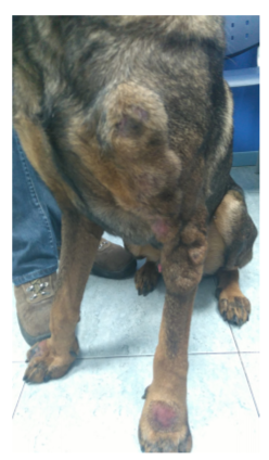
A 5-years-old intact male mixed breed dog was attended in our dermatology service with a 1-month history of nodular lesions. Multiple solid and non-pruritic nodules were located in face, trunk and limbs (Figures 1 and 2). No other symptoms were founded at clinical examination. Multiple biopsies of the nodular lesions were taken. The histopathologic findings were consistent with a CH. One month of treatment with tetracycline/niacinamide 500mg of each one twice daily and cyclosporine 5mg kg daily, was not effective. Administration of prednisone 1mg kg twice daily was able to resolve the skin lesions. After five weeks, adverse side effects occurred and prednisone was reduced at 1mg kg daily, and azathioprine (2 mg kg daily) was added. Lesions relapsed in one month and prednisone doses had to be increased. Three months later, dog development serious systemic symptoms and was euthanased.
Results
CH is a disorder of reactive dermal dendritic cells(2) . A dysregulation of immune response mechanism have been proposed(3) . Some immunomodulatory/ immunosuppressive drugs have been used in the management of CH. Tetracycline/niacinamide combination has proved to be successful(4). However, it was not effective in our patient that had to receive immunosuppressive doses of glucocorticoids +/- azathioprine to control the clinical course of lesions. An ongoing immunosuppressive therapy has also been reported to avoid relapses in some dogs(4), increasing the risk of development adverse side effects, as we saw in our case.
Conclusion
The management of canine CH can be complicated, especially in patients who need a continuously immunosuppressive therapy that can lead to a fatal outcome.
Figure 1.

Figure 2.
References
1. Affolter VK, Moore P. Canine cutaneous histiocytic diseases. In: Bonagura JD, ed. Kirk’s Current Veterinary Therapy XIII: Small Animal Practice. Philadelphia, PA: W.B. Saunders, 2000; 588-91
2. Affolter VK and Moore PF. Canine cutaneous and systemic histiocytosis: reactive histiocytosis of dermal dendritic cells. Am J Dermatopathol, 2000; 22: 40-8
3. Moore PF. A review of histiocytic diseases of dogs and cats. Vet Pathol, 2014; 51: 167-84
4. Palmeiro BS, Morris DO, Goldschmidt MH et al. Cutaneous reactive histiocytosis in dogs: a retrospective evaluation of 32 cases. Vet Derm, 2007; 18: 332-40
About
Copyright Statement
© All text and images in this publication are copyright protected and cannot be reproduced or copied in any way.Provided by:
Comments (0)
Ask the author
0 comments