menu sluit menu
Home Home
Login
Main navigation
  • Library
  • Calendar
  • e-Learning
  • News
    • Veterinary News In this section you find veterinary news
    • Recent Additions All content that was recently added to the IVIS library
  • Get involved
    • Donate Support IVIS, make a donation today
    • Media kit Promote your e-learning & events on IVIS
    • Add your e-learning & events to the IVIS calendar
    • Publish on IVIS Publish your work with us
  • About
    • Mission Our Mission Statement
    • What we do More info about IVIS and what we do
    • Who we are More info about the IVIS team
    • Authors See list of all IVIS authors and editors
  • Contact
User tools menu
User tools menu
Main navigation
  • Library
  • Calendar
  • e-Learning
  • News
    • Veterinary News In this section you find veterinary news
    • Recent Additions All content that was recently added to the IVIS library
  • Get involved
    • Donate Support IVIS, make a donation today
    • Media kit Promote your e-learning & events on IVIS
    • Add your e-learning & events to the IVIS calendar
    • Publish on IVIS Publish your work with us
  • About
    • Mission Our Mission Statement
    • What we do More info about IVIS and what we do
    • Who we are More info about the IVIS team
    • Authors See list of all IVIS authors and editors
  • Contact
Follow IVIS
  • Twitter
  • Facebook
Support IVIS

Breadcrumb

  1. Home
  2. Library
  3. European Veterinary Conference - Voorjaarsdagen
  4. EVC - Voorjaarsdagen - The Hague, 2017
  5. Cardiac surgery: what can you do in your practice?
European Veterinary Conference - Voorjaarsdagen
Back to Table of Contents
Add to My Library
Close
Would you like to add this to your library?

Get access to all handy features included in the IVIS website

  • Get unlimited access to books, proceedings and journals.
  • Get access to a global catalogue of meetings, on-site and online courses, webinars and educational videos.
  • Bookmark your favorite articles in My Library for future reading.
  • Save future meetings and courses in My Calendar and My e-Learning.
  • Ask authors questions and read what others have to say.
Sign in Register
Comments
Share:
  • Facebook
  • LinkedIn
  • Mail
  • Twitter

Cardiac surgery: what can you do in your practice?

Author(s):
Fossum T.W.
In: EVC - Voorjaarsdagen - The Hague, 2017 by European Veterinary Conference - Voorjaarsdagen
Updated:
JUL 05, 2017
Languages:
  • EN
Back to Table of Contents
Add to My Library
Close
Would you like to add this to your library?

Get access to all handy features included in the IVIS website

  • Get unlimited access to books, proceedings and journals.
  • Get access to a global catalogue of meetings, on-site and online courses, webinars and educational videos.
  • Bookmark your favorite articles in My Library for future reading.
  • Save future meetings and courses in My Calendar and My e-Learning.
  • Ask authors questions and read what others have to say.
Sign in Register
SHARE:
  • Facebook
  • LinkedIn
  • Mail
  • Twitter
    Read

    Cardiac surgery is one of the least commonly performed types of surgery in veterinary medicine. Open cardiac surgery necessitates that circulation be arrested during the procedure by inflow occlusion or cardiopulmonary bypass. Ligature placement using hand ties are useful in such situations and the ability to place hand-tied knots (vs. instrument tieing) should be considered a fundamental skill for cardiac surgeons. Secure knot tieing is critically important to successful cardiac surgery.

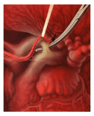
    Patent ductus arteriosus

    PDA is the most common congenital heart defect of dogs; it also occurs in cats. PDA causes a left-toright shunt that results in volume overload of the left ventricle and produces left ventricular dilation and hypertrophy. Progressive left ventricular dilation distends the mitral valve annulus causing secondary regurgitation and additional ventricular overload. This severe volume overload leads to left-sided congestive heart failure and pulmonary edema, usually within the first year of life. Atrial fibrillation may occur as a late sequela due to marked left atrial dilation.

    Surgical treatment

    Surgical correction of PDA is accomplished by ligation of the ductus arteriosus. Ligation of PDA is considered curative and should be performed as soon as possible after diagnosis. Secondary mitral regurgitation usually regresses after surgery due to reduction in left ventricular dilation. Inadvertent ductal rupture during dissection is the most serious complication associated with PDA repair. The risk of this complication decreases as the surgeon’s experience increases. Small ruptures, especially those on the back side of the ductus, often respond to gentle tamponade, but will enlarge and worsen if dissection is continued. Large ruptures must be controlled immediately with vascular clamps and then repaired with pledget-buttressed mattress sutures. Once bleeding is controlled, a decision must be made whether to continue surgery, or to abandon surgery in favor of repair at a later time. Second surgeries are more difficult due to adhesions at the surgical site, so complete occlusion should be attempted during the initial procedure, if possible. Often, simple ductal ligation is not possible after a rupture has occurred. In such instances, surgical alternatives include ductal closure with pledget-buttressed mattress sutures or ductal division and closure between vascular clamps. The divided ductal ends are closed with a continuous mattress suture oversewn with a simple continuous pattern. Ductal closure without division is safer than surgical division, but re-cannulation of the ductus may occur. Because ductal division requires added technical expertise, it should be undertaken only by experienced surgeons.
     

    Cardiac surgery

    Surgical technique

    Perform a left 4th space intercostal thoracotomy. Identify the left vagus nerve as it courses over the ductus arteriosus and isolate it using sharp dissection at the level of the ductus. Place a suture around the nerve and gently retract it. Isolate the ductus arteriosus by bluntly dissecting around it without opening the pericardial sac. Pass a right-angle forceps behind the ductus, parallel to its transverse plane, to isolate the caudal aspect of the ductus. Then, dissect the cranial aspect of the ductus by angling the forceps caudally approximately 45 degrees. Complete dissection of the ductus by passing forceps from medial to the ductus in a caudal to cranial direction. Grasp the suture with right-angle forceps. Slowly pull the suture beneath the ductus. If the suture does not slide easily around the ductus, do not force it. Regrasp the suture and repeat the process, being careful not to include surrounding soft tissues in the forceps. Pass a second suture using the same maneuver. Alternatively, the suture may be passed as a double loop and the suture cut so that you have 2 strands. Slowly tighten the suture closest to the aorta first. Then, tighten the remaining suture.

    [...]

    View full text
    Back to Table of Contents
    Add to My Library
    Close
    Would you like to add this to your library?

    Get access to all handy features included in the IVIS website

    • Get unlimited access to books, proceedings and journals.
    • Get access to a global catalogue of meetings, on-site and online courses, webinars and educational videos.
    • Bookmark your favorite articles in My Library for future reading.
    • Save future meetings and courses in My Calendar and My e-Learning.
    • Ask authors questions and read what others have to say.
    Sign in Register
    Comments (0)

    Ask the author

    0 comments
    Submit
    Close
    Would to like to further discuss this item?

    Get access to all handy features included in the IVIS website

    • Get unlimited access to books, proceedings and journals.
    • Get access to a global catalogue of meetings, on-site and online courses, webinars and educational videos.
    • Bookmark your favorite articles in My Library for future reading.
    • Save future meetings and courses in My Calendar and My e-Learning.
    • Ask authors questions and read what others have to say.
    Sign in Register
    About

    How to reference this publication (Harvard system)?

    Fossum, T. W. (2021) “Cardiac surgery: what can you do in your practice?”, EVC - Voorjaarsdagen - The Hague, 2017. Available at: https://www.ivis.org/library/evc/evc-voorjaarsdagen-hague-2017/cardiac-surgery-what-can-you-do-your-practice (Accessed: 30 March 2023).

    Author(s)

    • Fossum

      Fossum T.W.

      Professor of Veterinary Surgery, Vice President for Research and Strategic Initiatives
      DVM, MS, PhD, DACVS
      Department of Surgery, Faculty of Veterinary Medicine, Midwestern University
      Read more about this author

    Copyright Statement

    © All text and images in this publication are copyright protected and cannot be reproduced or copied in any way.
    Related Content

    Readers also viewed these publications

    • Journal Issue

      Veterinary Evidence - Vol 8 N°1, Jan-Mar 2023

      In: Veterinary Evidence
      MAR 19, 2023
    • Proceeding

      LAVC - Annual Conference - Lima, 2022

      By: Latin American Veterinary Conference
      MAR 18, 2023
    • Journal Issue

      Cirugía de urgencias - Argos N°246, Marzo 2023

      In: Argos
      MAR 10, 2023
    • Proceeding

      LAVC - Annual Conference - Lima, 2021

      By: Latin American Veterinary Conference
      FEB 19, 2023
    • Journal Issue

      Veterinary Evidence - Vol 7 N°4, Oct-Dec 2022

      In: Veterinary Evidence
      FEB 05, 2023
    • Journal Issue

      Patología cardiaca - Argos N°245, Enero/Febrero 2023

      In: Argos
      JAN 30, 2023
    • Proceeding

      SFT - Theriogenology Annual Conference - Bellevue, 2022

      By: Society for Theriogenology
      JAN 10, 2023
    • Journal Issue

      Urgencias y cuidados intensivos - Argos N°244, Diciembre 2022

      In: Argos
      DEC 31, 2022
    • Journal Issue

      Israel Journal of Veterinary Medicine - Vol. 77(4), Dec. 2022

      In: Israel Journal of Veterinary Medicine
      DEC 31, 2022
    • Proceeding

      ISCFR-EVSSAR Symposium - Italy 2022

      By: International Symposium on Canine and Feline Reproduction
      DEC 02, 2022
    • Journal Issue

      Patología endocrina - Argos N°243, Noviembre 2022

      In: Argos
      NOV 27, 2022
    • Proceeding

      ACVIM & ECEIM - Consensus Statements

      By: American College of Veterinary Internal Medicine
      NOV 11, 2022
    • Journal Issue

      Traumatología y neurología - Argos Nº242, Octubre 2022

      In: Argos
      NOV 10, 2022
    • Chapter

      Tibia and Tarsus

      In: Current Techniques in Small Animal Surgery (5th Edition)
      NOV 07, 2022
    • Chapter

      Femur and Stifle Joint

      In: Current Techniques in Small Animal Surgery (5th Edition)
      OCT 28, 2022
    • Journal Issue

      Medicina felina - Argos Nº241, Septiembre 2022

      In: Argos
      OCT 24, 2022
    • Chapter

      Sacroiliac Joint, Pelvis, and Hip Joint

      In: Current Techniques in Small Animal Surgery (5th Edition)
      OCT 17, 2022
    • Journal Issue

      Veterinary Evidence - Vol 7 N°2, Apr-Jun 2022

      In: Veterinary Evidence
      OCT 07, 2022
    • Journal Issue

      Veterinary Evidence - Vol 7 N°3, Jul-Sep 2022

      In: Veterinary Evidence
      OCT 04, 2022
    • Chapter

      Amputation of the Forelimb

      In: Current Techniques in Small Animal Surgery (5th Edition)
      OCT 02, 2022
    • Journal Issue

      Israel Journal of Veterinary Medicine - Vol. 77(3), Sep. 2022

      In: Israel Journal of Veterinary Medicine
      SEP 30, 2022
    • Chapter

      Carpus, Metacarpus, and Phalanges

      In: Current Techniques in Small Animal Surgery (5th Edition)
      SEP 26, 2022
    • Chapter

      Radius and Ulna

      In: Current Techniques in Small Animal Surgery (5th Edition)
      SEP 16, 2022
    • Chapter

      Humerus and Elbow Joint

      In: Current Techniques in Small Animal Surgery (5th Edition)
      SEP 10, 2022
    • Chapter

      Scapula and Shoulder Joint

      In: Current Techniques in Small Animal Surgery (5th Edition)
      SEP 05, 2022
    • Load more
    Provided by:
    Voorjaarsdagen Exit Party

    Every spring the European Veterinary Conference Voorjaarsdagen is organized. Important goals of the Voorjaarsdagen Conference are to build friendships between veterinarians at a national and international level, to enhance the quality and availability of veterinary medicine and surgery, and to foster the exchange of scientific information among veterinarians.

    Learn more
    Back To Top
    Become a member of IVIS and get access to all our resources
    Create an account
    Sign in
    Leading the way in providing veterinary information
    About IVIS
    • Mission
    • What we do
    • Who we are
    Need help?
    • Contact
    Follow IVIS
    • Twitter
    • Facebook
    International Veterinary Information Service (IVIS) is a not-for-profit organization established to provide information to veterinarians, veterinary students, technicians and animal health professionals worldwide using Internet technology.
    Support IVIS
    © 2023 International Veterinary Information Service
    • Disclaimer
    • Privacy Policy