
Pododermatitis (Bumblefoot): Diagnosis, Treatment, Resolution and Prevention
Author(s):
Updated:
FEB 24, 2024
Languages:
Read
Abstract
Pododermatitis is a disease of the integument of the plantar (bottom) surface of a bird’s foot. It can also spread to the dorsal (top) surface of the foot. It is caused by irritation, trauma, poor perching, or overweight. It will become progressively worse unless it is treated aggressively. There are seven stages of progression of the disease; the last is so severe that the bird’s foot might need to be amputated. Treatments consist of medical intervention in the early stages and surgical intervention in the later stages. The sooner the affliction is addressed, the more likely are the chances of full recovery.
Introduction
Pododermatitis, commonly known as “bumblefoot,” has become a frequently seen disease in companion and aviary birds. "Pododermatitis" is a general term for any inflammatory or degenerative condition of the avian foot. Many times birds will come into the clinician’s office for a routine annual examination, and the feet will show signs of subclinical or even more serious disease. The condition may range from very mild redness or swelling to chronic, deep-seated abscesses and bone destruction. If caught in the early stages, the underlying, predisposing factors may be corrected, and the disease will often be reversed.
1. Which species of birds are most at risk for developing pododermatitis?
Pododermatitis has been reported in many species of birds, but on a clinical level, it is particularly problematic in captive birds of prey, Galliformes (chickens and turkeys), Anseriformes (ducks, geese, and swans), waders, penguins, and many Psittaciformes (parrots). Of the psittacines, Amazons, budgerigars, and cockatiels are particularly vulnerable to this disease. The condition is frequently described in captive raptors, but it may occur in any avian species, including canaries and finches4. Because footpads are present in psittacines more so than in other species, birds in the parrot family are more likely to suffer from this disease.22
Figure 1. Pododermatitis at Stage 3 on the feet of a finch, side view (image courtesy Tamara Lowes; used with permission).

Figure 2. Pododermatitis at Stage 3 on the feet of a finch, plantar surface (image courtesy of Tamara Lowes; used with permission).

Figure 3. Extremely long nails cause the bird to stand in an unnatural position, leading to pododermatitis (image courtesy of Leila Marcucci, Bay Area Bird Hospital; used with permission).
The scales on the feet are composed of highly keratinized epidermal (the outermost layer of skin) tissue, and this tissue covers the lower leg (podotheca) and foot. The nails/claws are formed by plates of strong, keratinized tissue that enclose the terminal phalanx (last toe bone) of each digit. It is this keratinized tissue on the plantar surface of the feet that gets quickly worn away when the foot becomes irritated and sore.22
Birds most at risk for developing pododermatitis are obese birds having excess pressure placed on the feet; aged, sedentary and disabled birds; birds with limited mobility; chronically ill birds; and those with any kind of immune system weakness.21
1.1 What are the risk factors for developing pododermatitis?
- Previous foot or leg injury
- Hard, muddy, flooded, uneven, or rough floor surfaces
- Damp or unsanitary bedding litter
- Vitamin A deficiency
- Overweight
- Excessively dry skin
- Lack of activity
- Excessive activity due to fighting among flock members or guarding behavior
- Leg or conformation abnormality
- Improperly designed perches (plastic, sharp corners, incorrect diameter)
- Excessive accumulation of feces
- Poor diet
- Overgrown toenails
(Poultry DVM Bumblefoot in Chickens 2021 file:///L:/Pododermatitis/Bumblefoot%20in%20Chickens.html)
1.2 Causes or predisposing factors behind the development of bumblefoot
- Obesity and inactivity, which put more weight on the feet than it can handle. (Poultry DVM Bumblefoot in Chickens 2021 file:///L:/Pododermatitis/Bumblefoot%20in%20Chickens.html)
- Improperly designed perches: perches that are too small or too large and have no variety of diameter; those that are hard or uneven; dowel or hardwood surfaces; any rough-textured perches such as warming perches and all concrete perches, plastic perches, those covered in sandpaper or burlap, perches with sharp corners, perches that are too narrow, and perches or spirals made of sisal (J. Miesle)
- Hard, coarse floor surfaces, such as cement. These are common in aviaries, zoos, and breeding facilities. In poultry, floors may have hard, muddy, flooded, uneven, or rough surfaces. (Poultry DVM Bumblefoot in Chickens 2021 file:///L:/Pododermatitis/Bumblefoot%20in%20Chickens.html)
- Poor nutrition and Vitamin A deficiency. Birds need vitamins added to their food. (Do not put them in the water.) If they are on a pelleted diet, reduce and eliminate the pellets. Extra vitamins should not be given until the pellets are eliminated. They should be fed natural, non-pelleted diets consisting of fruits, vegetables, greens, some people foods, and seeds. Sunflower and safflower seeds may be given sparingly. They are high in fat and can lead to fatty liver disease. (J. Miesle)
- Poor husbandry: damp, unsanitary bedding and all substrates. An accumulation of feces and an overall unsanitary environment will lead to fungal and bacterial diseases and are a haven for parasites. Plain newspapers, paper towels, or other paper sources are the only things that should be used. (J. Miesle)
- Rope perches and natural wood perches wrapped in fleece or cohesive bandage tape to prevent sores are best. (J. Miesle)
- Overgrown toenails
- Stress, hypothyroidism,21 and hepatic (liver) dysfunction5
- Severe poxvirus lesions with secondary bacterial infections21
- Trauma, particularly among poultry:
-
Fighting among flock members,
-
Previous leg or foot injury leading to crippling
-
Frostbite injuries and thermal burns
-
Leg or conformation abnormalities
-
Cracks or worn-away areas and discoloration of the skin
-
Damage to the plantar surface of the foot. Injuries cause lesions to develop on the plantar surface of the phalanges or on the tarsometatarsus. Plantar decubital ulcers (pressure sores) are common. 20 (Poultry DVM Bumblefoot in Chickens 2021 file:///L:/Pododermatitis/Bumblefoot%20in%20Chickens.html)
-
-
Concurrent illnesses or conditions causing an abnormal standing position
- Arthritis. Pain in the joints causes the bird to walk on the sides of his feet; in this case, the toes bore most of the bird’s weight.
- Subdermal infiltrate swelling. The bird in Figure 4 suffered from mycoplasmosis as a result of a staphylococcus infection. He was unable to stand due to a nidus on the planter surface of the foot.
- Thickened pads on the bottom of the foot due to scar tissue or previous wounds which have been covered with these pads.
- Walking on the “heel” of the foot or the side of the foot due to swelling on the toes or center of the plantar surface of the foot. (J. Miesle)

Figure 4. After the nidus was resolved, the skin on the plantar surface barely covered the bones; this led to a pad of tissue developing. Because the pad was so large, he was forced to walk on the sides of his feet and toes. Arthritis set in because of his unusual limp and gait (image courtesy J. Miesle)
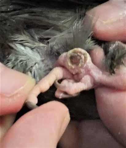
Figure 5. Subdermal infiltrate causing a nidus (pocket) of infection on the bottom of the foot, causing the bird to shift his weight to a toe and the tarsometatarsus (heel) (image courtesy J. Miesle).
2. Perches that are harmful to birds’ feet and leg joints
These perches are known to be detrimental to the plantar surface of the feet and cause pododermatitis in the feet and arthritis in the leg joints. Any of these can be wrapped with fleece or cohesive tape/bandage wrap to vary the diameter of the surface; however, most are round and need to be wrapped with high and low placement of the cushioned wrap (J. Miesle).
FLEECE: Purchase at least half a yard of fleece, and cut several 1-inch strips from it. Wrap the perch from the place closest to the cage bars, overlapping the fleece by half the strips. When you have reached the end of the perch, use a twist tie to wrap around the fleece and hold it on. It will need to be washed at least twice a week, so have extra strips ready to replace them. They wash well in the washing machine (J. Miesle).
Many people use Vetrap, but the author has found this tape to collect dirt very quickly, and it is so sticky many birds will not stand on it. Cohesive tape and fleece are better choices of products (J. Miesle).
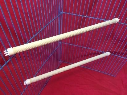
Figure 6. Plastic perches. These are usually too small for the bird, cause the bird to grip too tightly to stay balanced, and create pressure sores (image courtesy ebay.co.uk).

Figure 7. Kroger’s Tender Tape. This is a cohesive tape/bandage that wraps the perch well without being sticky. It will get dirty, so it must be changed at least once a week. A similar product can be purchased at the pharmacy with the name “cohesive tape” (image courtesy J. Miesle).

Figures 8. Rough-textured perches. These include cement, calcium, sandpaper, or warming perches. They irritate the plantar surface of the foot and cause sores and arthritis; they do nothing to keep nails short because the nails do not touch the perch (Fig. 8 image courtesy K&H Pet Products Bird Thermo-Perch). J. Miesle

Figure 9. Concrete perches should not be used (image courtesy Kathson Bird Perch Parrot Stand Cage Accessories Natural Wooden Stick Paw Grinding Rough-surfaced).

Figure 10. Sandpaper perch covers for dowel perches. Not only do they not keep the nails filed, but they also cause sores on the feet and arthritis in the legs. The owner can remove the sandpaper covers and wrap the perch with cohesive wrap or fleece, making sure to vary the diameter of the wrapped surface. Sandpaper covers also have a tendency to slip on the perch, causing the bird to grip too tightly to maintain balance, leading to arthritis (image courtesy Penn Plax Sanded Perch Covers for Small Birds) (J. Miesle).

Figure 11. Round dowel perches. These will place all the bird’s weight in the same place and are difficult for the bird to grip, forcing him to grip tighter all the time. Any perch that can become slippery puts additional strain on the muscles of the leg and foot to stay balanced (image courtesy Prevue Pet Products Birdie Basics Wood Perch 10 in) (J. Miesle).
2.1 Perches that are beneficial for the birds’ feet and legs.

Figure 12. Booda rope perches. Rope perches are available at some pet stores under a different name. These come in various diameters and lengths and also in spirals. They give the feet the soft, comfortable perching they need when they’re on their feet all the time and have minimal flying time. They must be removed and cleaned regularly so the bird is not standing on fecal material or chewing on it. When they become shredded, they must be replaced. When they become shredded, they must be replaced. Watch the bird carefully for signs it is chewing and possibly consuming the fibers on the perch. If he is doing that, remove the perch and replace with a wood perch covered in fleece (image courtesy JW Comfy Perches for Birds). (J. Miesle).

Figure 13. Hardwood perches, such as this manzanita perch, which offer a variety of diameters. Even these can be slippery, causing the bird to grip too tightly. And they are too hard for the bird's feet. They can be wrapped with cohesive tape or fleece strips (image courtesy Ebay). (J Miesle)

Figures 14. Flat wood perches.
Figure 15. Flat wire perches.
2.2 Covering perches made of wire and wood
Platform perches, wooden or made of wire. These come in a variety of sizes, shapes, and compositions. They can be found in wood, chrome, and coated wire. They should be covered with several layers of soft, padded material, such as flannel or fleece with a stack of paper towels under the fleece for comfort. (These coverings may be held down with binder clips for thinner perches or C-clamps for thicker ones.) (image 14 courtesy Pevor Wooden Parrot Bird Cage Perches, Amazon). (image 15 courtesy Platform Perches - Just for Pets). Never let any bird stand on open wire, as on grids on the bottom of the cage. Cover with paper towels or cloth towels. (J. Miesle).
3. Symptoms of bumblefoot in birds
- Dark, circular scabs on feet
- Redness, shininess, and small, red sores on the plantar surface of the foot
- Abrasions, cuts, tissue damage on the bottom of the foot
- Swelling and thickening of the skin
- Lameness and swollen joints in the feet or toes
- Reluctance to walk, stand, or grasp normally with one or both feet
- Ulcers on the soles of the feet2, 21
- Shifting of weight from one foot to the other
- Picking at the sores on the plantar surface of the feet

Figure 16. Healthy tissue in a cockatiel. Note that the nails are being kept short (image courtesy J. Miesle).
3.1 Means of infection
There are two ways infection can set it:
- Through a puncture in the skin of the base of the foot (a talon, a thorn, or a foreign object)
- Pressure sores (decubitus ulcers) on the bottom of the foot.9
3.2 Punctures to the skin leading to bacterial infections
Infections may occur when penetrations, such as cuts and sores, happen. Bacteria such as Staphylococcus aureus may enter the skin and cause damage if it has not been observed and treated. 2 Once the wound becomes serious, oral antibiotics, anti-inflammatories, and topical antibiotics will be needed.2 Celecoxib (Celebrex) is the best medication for birds for pain and inflammation (J. Miesle).
Systemic infections that result in decubital lesions or death can occur secondary to bumblefoot and are caused by virulent strains of S. aureus. S. aureus is frequently isolated from the lesions, but the birds will usually not respond to antibiotic therapy alone. These bacterial lesions may quickly lead to digital necrosis and gangrenous dermatitis. Staphylococci are by no means the only bacteria that might be recovered from diseased tissue: E. coli, Corynebacterium species, Pseudomonas species, and yeast are frequently cultured from the lesions.10
3.3 Decubitus ulcers
Decubital ulcers are open sores on the skin, often covering bony structures. Pressure sores occur because of uneven weight-bearing that leads to damage and devitalization of the skin. Both of these lead to bacterial and/or fungal infections of the skin. Once the process begins, a series of changes is initiated which will eventually damage the tendons of the foot and spread to muscles, joints, and other tissues. It can become a chronic disease, affecting the aortic and mitral valves of the heart and causing endocarditis (inflammation of the heart valves), vascular insufficiency (poor blood flow), lethargy, and dyspnea (labored breathing).9
4. Additional factors contributing to the development of pododermatitis
4.1 Malnutrition
In Psittaciformes and Passeriformes (songbirds), most lesions are believed to be the result of malnutrition. Poor nutrition causes the skin of the foot to become dry, flaky, and hyperkeratotic (developing a thick, outer layer of keratin on the skin). It is thought that dry, hyperkeratotic skin on the feet changes the mechanics of weight-bearing on the metatarsal pads. This condition is also precipitated by environmental deficiencies and systemic disease.4
Sunflower and safflower seeds have a high-fat content. Too many in the diet can lead to obesity in parrots and other pet birds. Traditionally, parrot diets have consisted of a mixture of seeds, with sunflower seeds being an important part of most diets (50% of the content of a sunflower seed is fat). Over the last decade, there has been an increase in the number of parrot owners that feed their pets commercial pellets; but this is also not a perfect alternative. Pelleted food contains more fat and protein than the amount most parrots need, and the oils added to the pellets (sometimes palm and coconut oils) may predispose birds to atherosclerosis (fat deposits in arteries). The heavier the bird, the more weight and pressure it puts on its feet, resulting in the development of pododermatitis. This is aggravated by a lack of flying; birds do not put pressure on their feet while they fly, so birds that do not fly are more predisposed to pododermatitis and obesity.16
The best diet for birds is one that is high In fruits, vegetables, greens, some people foods, and a moderate number of seeds (except sunflower and safflower. Seeds contain important Omega 3,6 fatty acids which protect the skin and internal organs. (J.Miesle)
See Appendix B, p. 59, for a list of foods that are nutritious for birds.
4.2 Vitamin A Deficiency
Some forms of pododermatitis are caused or exacerbated by Vitamin A deficiency. Birds that eat only seeds are susceptible to it since seeds are typically low in Vitamin A. This vitamin promotes appetite and digestion and also increases resistance to infection and to some parasites. The most obvious sign of a Vitamin A deficiency is a feather stain above the cere (the fleshy area which contains the nares or nostrils). The staining of the feathers above the cere reflects a discharge from the nostrils. Subtle differences may be seen as far as the color intensity of the cere and feathers and the overall condition of the plumage are concerned. A bird deficient in this vitamin may have pale, rough-looking feathers that lack luster. The cere may look rough instead of smooth, and you may see an accumulation of a yellow, dry scaling on the sides of the beak.2
Vitamin A deficiency weakens the epithelium (the thin, top layer of skin) of affected birds. Pressure sores, pressure ulcers, or decubitus ulcers occur when the bird is in the same position for a long period of time; the result is uneven weight-bearing. The ulcers are localized injuries to the skin and/or underlying tissues that usually blood flow to the soft tissue. The constant friction from the wrong types of perches can pull on blood vessels that feed the skin. Decubital ulceration on the plantar surface of the feet is common in older, obese, nutritionally deficient psittacines.20
4.3 Limited flying opportunities
Restricted flight opportunities lead to inactivity and obesity, and these produce excessive pressure on plantar surfaces, the toe pads, and the tarsometatarsus. Erosion occurs, and ulcers and staphylococcal infections develop.20

Figure 17: Grade I lesions. Note the shiny area on the foot pad (image courtesy Hari; used with permission).
4.4 Secondary disease
“Pododermatitis can also take place secondary to infectious or parasitic diseases, penetrating foot wounds, or leg injuries that affect normal gait and weight distribution. Epithelial damage that arises secondary to asymmetric weight-bearing on the metatarsal pad causes reduced circulation, microepithelial damage (microcysts; very small, round vesicles containing fluid and cellular debris), local impairment of the immune system, and ultimately, invasion of opportunistic pathogens.”19
For companion birds, raptors, and other wild birds, pathogenic bacteria introduced at traumatized sites may lead to abscessation (formation of abscesses), osteomyelitis (bone infection), or joint changes.9
(See Appendix A, p. 32, for more information on the treatment of raptors and case studies involving raptors,)
4.4.1 Arthritis
Septic arthritis may also play a part in the development of pododermatitis. Joints may become infected through a direct, penetrating wound or through the hematogenous route (spread through the bloodstream). Although the infection may be controlled, a decrease in the range of motion of the joint usually occurs.11

Figure 18: Grade 2 lesions (images courtesy Hagen Avicultural Research Institute; used with permission). Burgmann, Symptoms and treatment of bumblefoot.

Figure 19: Grade 3 lesions. Note that the bird is putting his weight on the outside of the foot, causing stress on that toe (images courtesy Hagen Avicultural Research Institute; used with permission). Burgmann, Symptoms and treatment of bumblefoot.
4.5 Contact with tobacco products
Many affected birds belong to cigarette smokers. Passive inhalation of cigarettes, cigars, pipe smoke, e-cigarettes, marijuana, and other types of airborne drugs not only causes ocular and respiratory disease in birds but also damages the integument. (Marijuana exposure also causes severe depression and regurgitation in birds and should be strictly avoided.) Pododermatitis has been observed in birds handled by smokers as the nicotine residues on the hands of smokers will cause this irritation. If minor, the lesions may spontaneously resolve when the client stops smoking or washes his hands and arms and changes his clothes before handling the bird.8
The feet and legs should be uniform in texture and color. The feet should have prominent scale patterns on the dorsal and plantar surfaces. Changes that result in the smoothing of the plantar foot surface can instigate chronic and severe foot and leg diseases. One of the common etiologies of foot abnormalities is contact with nicotine sulfate from the hands of cigarette smokers. The feet are particularly vulnerable to fungal diseases due to smoking toxicity.8
Pododermatitis has been observed in some birds handled by people who smoke routinely. Repeated exposure to the nicotine residues on the hands of smokers causes this local irritation. Macaws (and other birds with bare cheek patches) may suffer similar dermatitis on their cheek patches following repeated contact with a smoker’s hands.8
When toxic particulates and gases in the air assault a bird’s respiratory system, skin, and feathers, feather destruction, and plucking result. Heat causes the smoke to rise; then, when it cools, gravity brings it back down. The toxins land on the birds, their perches, their cage bars, toys, and food. It is also on the smoker’s hands, arms, clothes, and any other exposed skin— even the hair. If the smoker’s hands are coated with chemicals from holding the cigarette, it is easily transferred to his bird. In one case, an Amazon, who became very sensitive to chemical exposure, began mutilating his feet. The clinician determined the cause to be his perches which were covered with residue from cigarette smoke.14 Even if the smoker takes these precautions, his clothes, the furniture, rugs, curtains, and perches inside and outside the cage will be covered in smoke and ash. It is best for the smoker to stop smoking and have his home professionally cleaned (J. Miesle)

Figure 20. Grade 5 lesions on a cockatiel with arthritis (image courtesy of Julie Burge; used with permission). Note that there is fecal matter on the feet; this can cause additional infection in the already infected skin. (J. Miesle)
4.6 Contact with strong cleaners and disinfectants
These may also be responsible for irritation and dermatitis on the feet. Cleaning liquids should always be dry before the bird walks on the cleaned surface. Cleaning products should be carefully chosen so that chemical irritants are not used.8 Air freshener plug-ins and sprays should never be used around birds. (They also damage the respiratory system).14
4.7 Allergens
The allergy syndrome appears to be more common in the spring, suggesting a seasonal allergy. Seasonal recurrences of the lesions may be prevented by the oral administration of prednisolone about one month prior to the time that lesions typically occur.8
5. Avian Veterinary Examination
5.1 Physical exam
A bird’s skin is very delicate, and the skin of the foot reflects the condition of the rest of the dermis. The plantar surface of each foot should be checked daily by the owner, and the clinician should inspect the feet at the bird’s visits. Both owner and clinician should note the condition of the metatarsal and digital pads and look for loss of definition of the epidermis (seen as a shiny, reddened surface), swelling, erosions, ulcers, and scabs. Birds that are crippled from an injury or genetic defect are prone to developing sores on one or both feet due to the stance they must assume. The owner and practitioner must be observant for signs of pain or discomfort.6
If a bird is lame on one leg, he must bear his weight on the unaffected leg; this, in turn, can lead to pressure necrosis, infection, and subsequent pododermatitis on that foot as well. The clinician should always examine the other leg and foot closely when any lameness occurs. A complete examination, including radiographs, may require that the bird be anesthetized to reduce stress on it.6

Figure 21. Grade 4 bumblefoot in a parakeet (images courtesy Burgmann, Symptoms and treatment of bumblefoot, Hagen Avicultural Research Institute; used with permission).

Figure 22. Grade 6 pododermatitis in a parakeet (image courtesy Burgmann, Symptoms and treatment of bumblefoot, Hagen Avicultural Research Institute; used with permission).

Figure 23. Grade 5 pododermatitis in a cockatiel (image courtesy David Perpinon; used with permission).
5.2 Testing of the skin
Bacterial samples will be taken and evaluated at a microbiology lab in order to isolate the offending organism. An antibiotic sensitivity test may help determine the best course of antibiotic treatment to rid the bird of this condition or to manage it in the case of a chronic issue. A blood sample will be taken to check for other possible health conditions. X-rays will reveal signs of infection and will indicate any areas on the joints or bones where there’s been erosion or other damage.21
6. Clinical Grades of Pododermatitis
Hagen Avicultural Resource Institute definitions: The following classification grades have been lifted directly from the HARI institute website. (All are used with permission from HARI).
“A classification scheme grading from early clinical signs and progressing to severe lesions has been established. The clinical progression of the disease varies based on the species and the factors that contributed to the infection.”
Clinical Grades of Pododermatitis5
Grade I: Desquamation (shedding of the epithelial or skin tissue) of small areas of the plantar foot surfaces is represented clinically by the appearance of small, shiny pink areas and peeling or flaking of the skin on the legs and feet. Initial lesions are recognized as hyperemia (excessive amount of blood). Flattening of the skin of the digital and metatarsal pads is visible. These are the sites of maximum weight-bearing. (Thinning of the plantar surface of the foot with some reddening).
Grade II: These lesions progress if untreated, and bacteria invade the subcutis, resulting in a scab and mild swelling. (The subcutis is the deeper layer of the dermis, containing mostly fat and connective tissue). Smooth, thinly surfaced, circumscribed areas appear on the plantar metatarsal pads of one or both feet with the subcutaneous tissue almost visible through the translucent skin. No distinct ulcers are recognized. (The thinning of the plantar surface of the foot has progressed to the point that subcutaneous tissue such as tendons can be seen through the skin).
Grade III: The sores progress to form a caseous (having a cheese-like texture) abscess with marked swelling and pain. Ulceration of the plantar metatarsal pads occurs, and in some birds, a peripheral callus may form. Ulcers form on the soles of feet with callouses forming around the edges of the lesions. Some pain and mild lameness are present.
Grade IV: Infection of the tendon sheaths develops. Corresponding cellulitis tracks toward the intertarsal joint and the digits along with flexor tendon rupture. There is a necrotic plug of tissue present in the ulcer. Most species with ulcers and accumulation of necrotic debris exhibit pain and mild lameness. (Necrosis refers to cell death; the tissue turns black).
Grade V: Swelling and edema (cellulitis) of the tissues surround the necrotic debris. The digits of the foot may also be edematous (holding fluid). Necrotic debris starts to accumulate in the metatarsal area, indicating infection of the tendon sheaths. Severe lameness is common, and the entire metatarsal pad may be affected. This is generally a chronic lesion leading to osteoarthritis and septic arthritis of the tarsometatarsal-phalangeal joints. Cellulitis surrounds the area of necrosis, and the foot can be swollen with fluid. Tendon and metatarsal pads become infected; pain and severe lameness are present.
Grade VI: Necrotic tendons are recognized clinically as the digits swell and the flexor tendons rupture. Ankylosis and nonfunctioning digits usually present in recovery. (Ankylosis refers to the stiffness of a joint due to abnormal adhesion and rigidity of the bones of the joint, which is usually the result of injury or disease). (The digits are swollen and the necrotic flexor tendons on the plantar surface of the foot rupture. Even with treatment, non-functioning digits and joint fusion will be present).
Grade VII: Osteomyelitis develops. This is a bacterial bone infection leading to the destruction of the bone itself. (Bone infection can progress to systemic infection and death).

Figure 24: Comparison of a healthy plantar surface on the right and Grade VII severe case of pododermatitis on the left (image courtesy Lauren Thielen; used with permission).
6.1 Grading discussion
Grades I to III lesions are common in Psittaciformes and Passeriformes that are on all-seed or over-supplemented fruit and vegetable diets, are overweight, have no exposure to sunlight, or are kept on improper perches. With proper husbandry and nutrition, most cases recover with little medical intervention. The type of substrate and the size, shape and covering material of the perches may all influence the bird’s weight distribution on the toes and metatarsal pads. These affect the amount of skin wear on the plantar surface. For example, a perch that is too wide and flat may cause excessive weight-bearing on the toe pads, while one that is too small may cause excessive weight-bearing on the metatarsal pads.5
Early stages of pododermatitis show loss of the scale pattern on the foot, redness, and mild swelling. This damage can often be treated with topical softeners, improvement of the diet, and modification of the bird’s environment. Reversal is possible when the disease is at Grades I and II. The earlier the disease is caught, the more effective the treatment will be. The prognosis for the full recovery of lesions of Grade I to IV is usually more favorable than Grades V to VII lesions.5
Grade III bumblefoot is common in older, inactive birds that are fed inadequate diets. Early lesions (smoothing of the plantar foot surface) and hyperemia (an excess of blood in the vessels supplying an organ or other part of the body, Wikipedia) are frequently missed, and the birds are often not seen by the clinician until this stage. Some birds are not presented until they exhibit a non-weight-bearing lameness. This disease is dynamic and may move from one grade to the next quickly.5
Damage to the feet in Grades IV to VII usually requires surgical intervention since the changes that lead to infection also reduce the ability of antibiotics to be effective. Debridement of the wound, surgical removal of damaged tissues, and wound bandaging are treatments that are frequently used.5
Older budgerigars and cockatiels (5-10 years old) may have Grade V or VI lesions if precipitating factors are not corrected early. Bone changes and osteomyelitis may be present. Later stages are very serious and life-threatening. Since the development of antibiotic-impregnated beads, veterinarians can treat later stages more successfully than they could before. Amputation is sometimes necessary. Prosthetics may be used for birds such as ducks that cannot survive with only one good foot.5
7. Treatments for pododermatitis
Perching, cleanliness, and nutrition need to be addressed.
- Below you will find information on perching.
- The cage must be kept scrupulously clean with an antibacterial cleaner once a week or more often if it needs it. Clean all areas daily and watch for droppings and food debris as these need to be cleaned constantly.
- Nutrition: If the bird is on pellets, reduce and replace them with fresh fruits, vegetables, some seeds (not sunflower or safflower as these are high in fat and can lead to fatty liver disease), people foods, seed supplements.
See Appendix B, p. 41, for a list of healthy foods that may be added to the current diet.
If the owner does not have an avian veterinarian, he may utilize the following instructions. If he does have an avian veterinarian, these instructions may be used in addition to those given by the avian veterinarian.
7.1 Treatment of the lesions, Grades 1 and 2
1. Take the bird to the veterinarian immediately. He will need to determine the extent of the damage to the feet and guide you in their healing. If the veterinarian wraps the feet, find out how often you need to bring him back to have them treated and rewrapped, or ask him to let you change the bandages there so you can do it at home. (If you do not have an avian vet, contact the author so she can help you find one). Follow the rest of the recommendations below. Do not use a heating pad! Get a prescription for Celebrex (celecoxib). Do not use meloxicam. It is not as effective and can cause digestive problems.
2. Change all perches to soft rope perches or fleece-covered natural wood perches immediately. Be careful to get the right diameter of perch for your species of bird. Until you can get them changed, buy at least 1 yard of very soft fleece. Wrap all perches with overlapping, 1-inch strips of fleece held down by short pieces of twist ties on the end that the bird does not stand on. Once you get the rope perches, wrap them with the fleece, or continue using natural wood perches wrapped in fleece. You can wrap any rough-textured perches, dowel perches, or warming perches with fleece until you can replace them with rope or natural perches. Remove all rough textured, plastic, dowel, and warming perches. Platform perches must be covered in at least 2 layers of fleece held down by binder clips. The perch will be larger with the addition of the fleece. Keep the fleece on indefinitely.
See 1.1 for descriptions of perches that should not be used for any birds, but especially the bird with sore feet.
3. Clean the feet with antibacterial soap and rinse and dry well; apply and massage in creams. There are two prescription creams that you can get from your avian veterinarian: Gentamicin Sulfate Cream (©Perigo or generic and Neomycin and Polymyxin B Sulfates and Bacitracin Zinc Ophthalmic Ointment, USP (©Bausch +Lomb). These may be alternated until you find which works better.
Alternate or additional topical therapy for Grades 1 and 2 if the prescription creams cannot be purchased: Get Aloe Vera Gel from Walgreens (or a similar gel). Put a small amount in a small container, and add a little distilled water until you get a medium-gel consistency. Begin to massage the feet with a small amount of triple antibiotic ointment, such as Neosporin, followed by the Aloe Vera Gel. Do this treatment two or three times a day. It is available online from Walgreens. Do not use Aloe Vera plant liquid. It is not sterile and is not as effective as the gel.
4. Keep the bird on fleece-covered perches for a long time, at least until the feet are completely healed and possibly longer, since some birds have a relapse of the condition. Make sure you remove the fleece strips twice a week and replace with clean ones. They are washable.
5. If the bird shows discomfort, place him in a storage bin on several soft towels until his condition improves. Put his food, water, and toys in the bin with him. If he’s flighted, cover with a framed piece of aluminum screening or some other type of open covering. When he’s out of the cage or bin, keep him on soft, clean surfaces at all times. You can put soft towels on the surfaces the bird walks on, such as the top of the cage or on any play areas.
7.2 Additional information concerning treatment for Grades 1 and 2
Grade I and II lesions generally respond to keeping the foot clean, applying the medications, and correcting underlying management or nutritional deficiencies. With waterfowl, this frequently involves changing the dimension, shape, and surface of the enclosure, adding adequate swimming areas, and keeping everything very clean.20
Less severe cases may be treated with a combination of environmental and medical methods. Correction of the underlying predisposing factors will often reverse this disease process. Change in diet, changes in perches, frequent exposure to sunlight, improved cleanliness, environmental changes, and the application of a topical antibiotic or antimicrobial cream containing steroids often resolve the problem. (See 7.1 for medications used) These should be used with caution to prevent toxicity. Many topical products are available, such as softening agents for dry, scaly feet, topical antibacterial creams for acute inflammation and swelling and ointments for granulating wounds. (Granulation tissue is new connective tissue and microscopic blood vessels that form on the surfaces of a wound during the healing process [Wikipedia]).20
The addition of ©Vetomega is recommended to aid in healing the wounded skin tissue. (Link to purchase of VetOmega from Scott Echols.)
For all levels of disease, the owner must be willing to continue with the treatment at home, possibly for months. He needs to be made aware that some individuals are highly susceptible to recurrences. If the bird does not respond to these changes, or the condition continues to deteriorate, more complex medical treatment will be necessary.20 At that point, you must consult a veterinarian.
It is of greatest importance to discuss all of the above treatments with your avian veterinarian.
7.3 Treatment of the lesions, Grades 3
1. Take the bird to the veterinarian immediately. He will need to determine the extent of the damage to the feet and guide you in their healing. If the vet wraps the feet, find out how often you need to bring him back to have them treated and rewrapped, or ask him to let you apply the cream or ointment and change the bandages there so you can do it at home.
If you do not have an avian vet, contact the author so she can help you find one, and follow the rest of the instructions below.
2. Prepare the lesions for treatment at home
To prepare the lesions for the topical antibiotics, they need to be thoroughly cleaned. Washing the feet with antibacterial soap is sufficient for Grades 1 and 2, but for Grades 3-7, preparation is more comprehensive. One recommendation involves soaking the affected foot/feet in a shallow dish filled with warm water with Epsom Salts added to it. The recommended strength is about one teaspoon of Epsom Salts mixed in with one gallon of clean, warm drinking water. The Epsom Salts will draw out any toxins. Soak for as long as the bird will tolerate it, at least 5 minutes per foot. Do this twice a day. Soaking the feet will soften and remove any scab, which allows the lesion to drain any pus and debris. Do NOT remove the scab or other material without soaking it first. Allow the material to fall off itself. Finally, flush the cavity with diluted hydrogen peroxide to clean the wound out and destroy any bacteria. 2 (Hydrogen peroxide should be diluted at a 1:10 ratio with water. One part HP, 9 parts water.)
3. Transfer the bird to a storage bin of the appropriate size for his species. Prepare the bin by laying down at least 2 soft towels on the bottom. While the bird’s feet are healing, buy at least 1 yard of very soft fleece. Wrap all perches with overlapping, 1-inch strips of fleece held down by a short piece of twist tie at the end that the bird does not stand on. Make sure perches are the appropriate size for your bird’s species. Cut a piece of fleece (or two smaller pieces) to fit the bin. Make extra pieces to replace the large pieces when soiled. They are washable. Place the bird’s food, water, and toys in the bin. Do not use a heating pad!!
4. Clean the feet with antibacterial soap and rinse and dry well; apply and massage in creams. There are two prescription creams that you can get from your avian veterinarian: Gentamicin Sulfate Cream (©Perigo or generic ©G&W, and (©Neomycin and Polymyxin B Sulfates and Bacitracin Zinc Ophthalmic Ointment, (USP Bausch +Lomb). These may be alternated until you find which works better.
Follow the information in 7.1 and 7.1.1. Obtain VetOmega as discussed in 7.1.1. Purchase the two creams under 7.1.1, #3. If you cannot get the prescriptions for these, use the OTC medications in the following paragraph. Get the Aloe Vera Gel from Walgreens and mix a very small amount with distilled water to make a medium-thin gel.
Begin to massage the feet with a small amount of triple antibiotic ointment, such as Neosporin followed by the Aloe Vera Gel. Use only this gel or a similar brand. Do this treatment 3 times a day. You can reduce this to 2 times a day as the skin heals. It is available in-store and online from Walgreens. Do not use Aloe Vera plant liquid. It is not sterile and not as effective.
5. While the foot sores are healing and the bird is in the bin, change all perches to soft rope perches or wrap the perches in fleece strips. Be careful to get the right diameter of perch for your species of bird. Have the cage ready for the bird by the time he is ready to go back into it. Do not wait.
6. Once the feet have healed, the bird may be returned to his cage. He will have to be on fleece for a long time and possibly indefinitely since some birds have a relapse of the condition due to the skin having been compromised. Make sure you remove the fleece strips twice weekly and replace them with clean ones. You will eventually be able to change them just once a week. Replace the fleece weekly if there are no open sores, blood or fluid coming from the wounds. Replace the fleece daily if there are any open sores, blood, or fluid on the feet.
7.3.1. Dressings and bandaging
The severity, type, and path of infection will direct treatment. No matter what, the owner should keep the bird’s feet sanitized, and tailor the living environment to both promote healing and to eliminate destructive perches and surfaces.21 (Installing rope perches and wrapping all perches with fleece are the best way to provide soft surfaces). Oral antibiotics and antibiotic ointment will control the infection. Bandaging may be recommended in order to reduce the opportunity for pathogens to enter the wounds. In more severe cases, surgery (including debridement of abscesses) will help to save the feet, and life, of the bird. Antibiotics will be essential in resolving infection, and the bird can be kept comfortable with pain and anti-inflammatory medication. (Celecoxib [Celebrex] is the best anti-inflammatory and pain medication for birds; do not accept meloxicam as it is not meant for birds and is not as effective as celecoxib. It also has gastrointestinal side effects that celecoxib does not have.) 21
Medical therapies include the application of topical agents in order to toughen the plantar skin of the foot. Softening agents are also helpful in cases in which the epithelium has become thin. Antimicrobial and anti-inflammatory creams and ointments and hypertonic footbaths provide infection and inflammation control. (Hypertonic refers to a solution with higher osmotic pressure than another solution. If a cell is placed in a hypertonic solution, water will leave the cell, and the cell will shrink [Merriam-Webster.]) 5
Once the wound is clean, apply liberal amounts of antibiotic ointment as prescribed by the veterinarian, and then carefully wrap the foot or feet with gauze; this will keep the cavity clean and the ointment in place and prevent the bird from chewing on it. If the pododermatitis is in the very severe category, a ball bandage may need to be adhered to the foot. The lesion needs to be cleaned and ointments reapplied twice a day until the feet appear healthy.2

Figure 25. A ball bandage is one option for the management of a closed-toe fracture (image courtesy Dr. Ariana Finkelstein, Lafebervet.)
6. In severe cases, a hydroactive dressing may be used (hydroactive: the act or process of combining or treating with water: As the introduction of additional fluid into the body, hydration sometimes helps to reduce the concentration of toxic substances in the tissues dressing and can be used to facilitate healing of the wounds [Merriam –Webster]). Hydrophilic dressings mix easily with water. Moisture-vapor permeable dressings or hydrocolloids (a substance that forms a gel when mixed with water) dressings should be applied topically to enhance wound healing for open, granulating wounds or post-operative incisions. Applying a topical antibiotic and bandaging the feet with sufficient padding to reduce and better distribute pressure on the plantar surfaces is required in many cases. The types of bandaging methods may include simple toe bandages, interdigitating (between the toes) bandages, and ball bandages. Bandaging of affected tissues may go on for several months until the bird responds to the new diet and environmental deficits have been corrected.5
Initially, the bandage may require daily changing. The frequency of bandage changes can be reduced as the wound becomes less exudative (producing fluid, especially in inflamed tissue). Once granulation tissue forms at the edge of the ulcers, scabs should be removed by the clinician, and the lesions should be kept clean to facilitate healing.5
7.4 Additional therapy for lesions, Grade IV to VII
- Cleansing and treating the wounds daily with multimodal medications and changing the bandages daily
- Treating the wounds locally by using the antibiotic-impregnated matrix; antibiotic-impregnated beads are implanted into the wound
- Administering systemic antibiotics parenterally (other than by mouth) in the beginning followed by oral dosing
- Prescribing anti-inflammatory analgesics to provide pain relief. NSAIDs (non-steroidal anti-inflammatory drugs; e.g., celecoxib) or synthetic opioids may be used as needed.
- Testing for systemic infection in more advanced cases of decubital ulceration (bedsores or pressure sores)
- Performing a complete blood count.
- Taking swabs regularly from within the abscesses for fungal and bacterial cultures. E. Coli, Staphylococcus, and Candida albicans are commonly isolated pathogens.5
It is of greatest importance to discuss all of the above treatments with your avian veterinarian.
7.5 Goals for treatment of advanced pododermatitis, Grades IV To VII
Advanced bumblefoot is diagnosed from the bird having a necrotizing abscess on the plantar surface of the foot. Depending on the location and chronicity of the abscess, infection may or may not extend to neighboring joints, tendon sheaths, and bones.10
The goals of advanced bumblefoot treatment are:
- To reduce infection, inflammation, and swelling
- To establish drainage as needed
- To begin antibacterial topical and oral therapy to eliminate underlying pathogens
- To manage the wounds in order to promote rapid healing
- To initiate surgical intervention when needed
- To ensure an adequate diet, and
- To address environmental deficiencies.5

Figure 26. Grade VI Pododermatitis in a four-year-old budgerigar with a straight perch in his cage for years (image courtesy of K. Gerbaga Özsemir; used with permission).
This will be accomplished by:
- Correcting any perching and bedding issues
- Adjusting nutrition as needed
- Correcting potential husbandry issues; cleaning and disinfecting the bird’s enclosure in order to eliminate injuries and stresses associated with those
- Correcting the bird’s diet when it is found to be lacking sufficient Vitamin A. Vitamin A injections are an option. Recommendations will be made by your avian vet or through research to supplement the diet to increase vitamin A.
- Addressing bacterial infections, whether localized in the wound area or systemic. Cephalexin antibiotics have been used successfully in cases that involve infection that has spread through the body.
- Increasing exercise and adjusting the diet of obese birds
- Treating and dressing any wounds or ulcerations that are present to encourage healing
- Using surgical treatments, natural healing products, therapeutic lasers, and even acupuncture as needed. These are determined by the extent to which the pododermatitis has progressed and the recommendation of the avian veterinarian.21
It is of greatest importance to discuss all of the above treatments with your avian veterinarian.

Figure 27: Grade VII bacterial pododermatitis. This lesion usually develops following pressure necrosis with a subsequent bacterial infection (image courtesy Harrison: Clinical Avian Medicine, 2006).

Figure 28. The nidus pocket from Figure 5. The scab that resulted after treatment is covering the nidus hole. The pocket is deep under the thin scab and will bleed if the scab is removed. The nidus goes down to the bone and is the result of a staphylococcus infection caused by mycoplasmosis synoviae. Infections and diseases can cause swelling in the feet resulting in pododermatitis (image courtesy J. Miesle).
7.6 Additional treatment of advanced cases
Advanced cases may warrant surgical debridement (cleaning and cutting away of dead tissue) of fibrotic and exudative material as well as attempts to close the wound with sutures. Debridement should be approached cautiously since hemorrhaging can occur from decubitus. Surgical excision of the abscess or amputation of a severely traumatized digit may be indicated.5
Surgery is often necessary to repair damage to the tendons and ligaments. This is a long, slow process, and it may take months before the feet are healed. Even after healing is complete, the foot may still be tender for several weeks. Preventing trauma and maintaining the patient on a soft footing is important to avoid recurrence. Waterfowl should be returned to the water as soon as possible to prevent further damage.5
Treatment for Grade IV should include drainage, irrigation, and closing of the wound when the infection has been resolved. The prognosis is fair. Treatment for Grade V to VII lesions must be vigorous, and the prognosis is guarded.5
(See Appendix C, p. 44, for more information on laser treatments for pododermatitis,)
8. Consequences of neglecting to provide treatment for pododermatitis
If the bird owner notices the formation of these sores, initiates veterinary treatment, and makes positive changes to the bird’s living environment, the prognosis for healing is good. However, without veterinary attention and environmental improvements, the sores typically turn into painful abscesses, which enable opportunistic pathogens (usually S. aureus) to breach the surface of the thinning skin.21
8.1 Pain, arthritis, and infection
The pain from these lesions causes increased weight-bearing on the unaffected foot, forcing the bird to bear its weight disproportionately. As a result, many birds suffer from bilateral pododermatitis. The plantar location of the lesion is constantly under forces of pressure, movement, and contusion (bruising); in addition, the bird’s feet are constantly exposed to contaminants.9 These birds are prone to arthritis as well, and this disease only worsens with time.
Celecoxib should be provided for any birds experiencing pain and inflammation for any reason. It is far superior to Meloxicam and has fewer side effects.

Figures 29 “Infectious pododermatitis with gross swelling of a foot in a snowy owl. The central scab was removed, and a large amount of liquid pus was present within the foot.” Figure 30. “After debridement and application of a topical ointment and dressing, an interdigital bandage was applied as well as a custom-fitted silicone shoe” (image and text courtesy B. Speer: Current Therapy in Avian Medicine and Surgery).
8.2 Necrosis, lameness, and decreased quality of life
In due time, the infection encroaches upon joints in the feet and bones in the legs, and surrounding tissues become necrotic. Ulcers may form on the feet, and the bird may become progressively lame.21 “Birds beset by advanced and untreated bumblefoot can become so systemically infected that their lives are unsustainable.” 21 If left untreated, the lesions lead to crippling deformities, sepsis, and poor quality of life.9 Bacterial infections that begin in the pads of the foot can ultimately lead to a bird’s death. Many surviving birds endure chronic abscesses and the amputation of a leg.21 Unless the condition is treated, the infection will eventually eat into the bone and travel to other parts of the body. This is a painful condition that can lead to death.21
8.3 Osteomyelitis involvement
If systemic infection and pain can be controlled, the above therapy may be attempted. If the disease state becomes extreme, osteomyelitis occurs, and the prognosis for recovery decreases dramatically. The owner must be forewarned that the therapy will be of a long duration, and the prognosis is poor. The owner and practitioner will need to discuss the ethics of such long, continuous treatment when the degree of disease is so advanced that the bird cannot stand without severe pain. Euthanasia will need to be considered under such circumstances.20
8.4 Limb Amputation
If a bird has had a pelvic limb amputation, it is possible that pododermatitis will develop in the opposite leg. These birds benefit greatly from having soft, wide, padded perches and platforms provided for them. It is also possible that the wounds will be so severe that they do not respond to medical or surgical therapy, and the second foot or leg will need to be amputated. Euthanasia must be discussed if that occurs.6
9. Prevention of pododermatitis
Pododermatitis is easier to prevent than to treat. Bumblefoot may be prevented by taking the proper steps to ensure that the bird’s living conditions are correctly designed and it is given a nutritious diet. Prevention of pododermatitis involves constant vigilance for early signs of hyperkeratosis, baldness, flaking of the skin of feet and legs, redness or swelling. Early correction of the underlying causes will avert future severe disease.19
9.1 Choosing the right size cage
Cages should be large enough to accommodate several different types of perches. Cages should contain horizontal bars for climbing; these will help prevent trauma to the foot pads from vertical bars which need to be gripped to slide down. Care should be taken to make sure that the wire is smooth and contains no sharp places which could puncture or scratch the feet. Even powder-coated cages may contain sharp points which could damage the feet. Any rough places should be looked for and smoothed over with a file.19
For birds housed in wire enclosures, the walls of the enclosure should be designed with vertical bars or solid barriers to minimize the tendency for hanging from the wire. Never house birds in galvanized wire structures. Birds will chew on that type of wire and fall ill with metal toxicosis. Selection of proper perch size, shape, and covering for a particular species of bird is very important. 19 (The reader may contact the author for more information.)
If a bird's case of pododermatitis is severe, it would be helpful to house him in a “flat,” wide cage. (The term “flat” is used by manufacturers and sellers to distinguish it from the square and tall cages.) These are especially nice for handicapped, aged, or ill birds, not just for those with bumblefoot. Yaheetch® and Chewy® carry these cages, among others.

Figure 31. The author’s bird in a bin with towels layered for softness. When arthritis becomes very painful, or painful foot sores are present, placing the bird in a bin on towels is preferable to keep him in the cage. There are no bars, multiple towels will protect the feet, and the bird will move about much more easily. Food, water, and toys may be placed in the bin with the bird. For bumblefoot, the additional heat from a heating pad is contraindicated because it will aggravate the condition, making any inflammation, infection, or irritation worse.
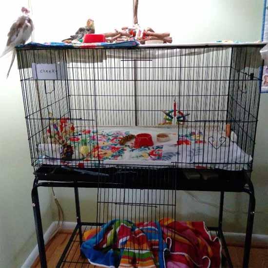
Figure 32. A flat or wide cage. As you can see, toys can be placed on the top, The food and water dishes can be placed close to the bird’s favorite spot—in this case, the front right corner. There are several larger pieces of fleece there for extra softness. And there is a small perch on the left front corner, close to the floor of the cage. The bird can stand on that if he wishes. It is wrapped in fleece. There are 3 soft towels on the floor of the cage and toys on the sides and back.
9.2 Choosing the correct perches
The perches should be of various textures and sizes so the bird is not standing on the same plantar surface all the time. They should be of different shapes: oval or flat are better than round ones all of the same size. Rough-textured perches, such as concrete and sandpaper should NEVER be used since they will upbraid the tissue of the foot. (Perches that are made of plastic and dowel wood, perches that are too small or too large, warming perches, and those that have no variance in size, should not be used for any bird.)1 (See figures in Section 2 for descriptions of poorly designed perches.)
Perches that are too smooth, such as dowels and plastic perches, will force the bird to grasp tighter all the time, causing pain, and eventually, arthritis in the feet and legs; in addition, the bird can easily fall, causing bone breaks or wing damage. They should vary in diameter and offer the bird alternating resting places for the plantar surface of the foot. Rope perches (such as the Booda perches) come in a variety of sizes, and along with natural tree branch perches, grape wood perches, and platform perches, provide a good standing surface. Platform perches are very comfortable for birds. They should be covered with several layers of soft material, such as flannel or fleece; these may be anchored down with binder clips or small clamps. (J. Miesle)
You can purchase flannel fabric from any fabric store or online and cut several layers to fit the platform. Do not use paper towels or other fabric due to their rough texture; in addition, both could be easily ingested. If the sores are not too bad, loose paper towels may be placed on top of the secured ones for easy cleanup. These cloth towels must be laundered daily or whenever soiled, so you will need several. A half-yard of fleece will give you many pieces to work with. (J. Miesle)

Figure 33. Stage VI pododermatitis; These sores are necrotic and oozing (image courtesy Brisbane Bird Vet; used with permission).
Perches that are both too hard, such as Manzanita perches, and too smooth (such as dowel and plastic perches), sharp-cornered perches, rough pedicure perches, sandpaper and rough-textured/concrete perches should be removed and replaced with rope perches and natural wood perches. Hard perches should be wrapped with a cohesive material such as Kroger’s Tender Tape® or other cohesive tape which can be purchased at a drug store, or one-inch strips of flannel. This will provide both padding and changes in diameter when the material is wrapped at varying intervals and thicknesses. Natural perches with different circumferences and textures are good but should be covered with cohesive tape or fleece strips. Birds should be encouraged to perch in different places and varying surfaces. This can be achieved by placing food and water dishes in different areas and changing the position of the favored perches.2 Birds will choose a favorite place within the cage, so whatever perch is there is the one they will perch on (J. Miesle).

Figure 34. Grade VII infectious pododermatitis in a chicken; This bird’s staphylococcus infection is destroying the skin, making it impossible for the skin to be sewn together (image courtesy Farmer’s Weekly).
(Some people think birds choose a favorite perch when in reality they are choosing a preferred space in the cage. Take note of that and put the softest perches there).
9.3 Choosing correct cage and enclosure flooring
These can be a problem. The wire should never be the only thing that any bird stands on, whether it be the floor of the cage or aviary. It can damage the feet and cause cuts and bruises. Hard or wire flooring that the bird with pododermatitis—and any bird-- stands on should be covered with soft towels to protect the feet, facilitate a more comfortable surface on the floor, and speed the healing process. The grids on the bottoms of the cages of a bird with bumblefoot should be covered with two layers of soft towels without anything on top of them, even paper towels. The grids on the bottoms of non-affected birds’ cages should be covered with either a towel covered with paper towels or just several layers of paper towels. Birds should never stand on grids or wire. Any surfaces an affected bird touches should be carefully sanitized and kept clean, and it should always be very soft (J. Miesle).
9.4. Sanitation and substrates
Proper hygiene is of the utmost importance in preventing bumblefoot.
- Cages and perches need to be cleaned and disinfected daily to avoid contamination from fecal matter and bacterial growth on surfaces and in food.
- Soft foods should be removed within two hours to prevent bacterial growth.
- Wipe down any surface that fresh foods have been on as soon as the food has been removed to prevent bacterial growth
- Seed, food, and water cups should be checked frequently during the day for droppings.
- Food and water cups need to be cleaned daily and replaced with fresh food and water as needed.
- The grates and trays of the cage must be cleaned daily since many birds spend time on the bottom of their cages where they may come in contact with the droppings. Paper towels on the grates (or trays if there are no grates) will make clean-up easier.
- All play areas must be kept very clean. Any play surface is a potential source of bacterial and fungal pathogens that could invade the surface of the bird’s feet.
- T-stands, toys, and anything the bird encounters or stands on must be kept scrupulously clean. Droppings, regurgitation, and soft foods should be cleaned up immediately to prevent consumption, reinfection, and transmission of disease.
- Care must be taken to choose a cleaning product that will not harm the healthy or damaged tissue. All cleaning products need to be dry before allowing the bird to stand on them.3 White vinegar (2 ¼ cups to a gallon of water) is a good cleaning liquid.
- Do not use substrates on the bottom of the cages. These include nut shells, wood chips, grains, corn husks, moss, pine cones, and bedding made for reptiles and small mammals—any type of bedding. These not only provide opportunities for bacterial and fungal spores to grow, but they also put dust into the air that harms the bird’s breathing and make it impossible for the owner to observe the droppings. Use plain newspaper, and place it where the bird cannot reach it.
9.5 Proper Nutrition
Nutrition is extremely important. Many affected birds are primarily seed-eaters. Feeding a balanced diet of some seeds (very few sunflower and safflower seeds since they are high in fat and can lead to fatty liver disease), fresh fruits and vegetables, greens, and some human foods are critical, as is providing fresh water for bathing and drinking. Proper nutrition often will prevent or even reverse early bumblefoot in Psittaciformes. The diet should be corrected to promote needed weight loss for obese birds and to increase general nutritional balance, with emphasis on the replacement of Vitamin A precursors.13,20 Providing Vitamin A injections if needed and foods high in Vitamin A will prevent more damage to the feet.
9.5.1 The harmful effects of excess protein
Excess protein when stored in the body promotes the growth of internal bacteria which are excreted through the skin. In areas where there are feathers, those feathers will usually absorb the protein. In bare areas, such as the feet, these bacteria will present themselves as pink, red, and then blue "calluses." These most often show up on the bottom of the feet; however, may also appear on the top or on the tips of the toes, above or under the bird's toenails. It is important to reduce the protein in the bird's diet to stop the progression of this condition. 2
9.5.2 The benefits of vitamins, minerals, and supplements
Birds require supplemental vitamins and minerals to aid in the prevention of bumblefoot and other diseases. Birds require Vitamin A and biotin (a B-complex vitamin) supplementation to ensure healthy skin development. Deficiencies in these vitamins may result in pododermatitis and focal hyperkeratosis (plantar corns). Bird owners should provide multivitamins, minerals, and essential fatty acids/amino acids supplementation to prevent these diseases.12
For the affected bird with Stages III to VII, initial injections of Vitamins A, B-complex vitamins, and Vitamin D3 are advised, along with continued use of oral supplementation of multivitamin/mineral/essential fatty acid/amino acid preparations. Your avian veterinarian may also want to provide those injections to birds with Stages I and II. 1,12,16 ©Avi-era Vitamins and ©Missing Link minerals are excellent choices for vitamin and mineral supplements. They should be given daily, and alternated, in small amounts on the food, not in the water. Omega 3 and 6 essential fatty acids may be added in the form of ©VetOmega, available through your avian veterinarian or directly from Dr. Scott Echols. This product provides your bird with the oils it needs for healthy skin and feathers and protects the internal organs. This link will allow you to order it yourself if your veterinarian does not carry it. Encourage him to begin to offer it to his clients. (J. Miesle)
https://www.vetomega.com/?fbclid=IwAR330QUDMPrtHY-rzR6AMrMg- vbPWPf_SmcfmA36qt_q4LpjpM5t0K0wYuAet

Figure 35. Padding perches with foam helps prevent bumblefoot (images courtesy Hagen Avicultural Research Institute; used with permission). Burgmann, Symptoms and Treatment of Bumblefoot.
9.6. Exercise
Exercise will aid in preventing pododermatitis and healing from it since he will spend less time on his feet if allowed to fly. Make sure everything he stands on is protected with soft towels! Allow the bird out-of-cage time frequently throughout the day so that it can fly and strengthen its legs; this will take the pressure off the feet. Exercise will also aid in lowering the obese bird’s weight.11 If the bird does not fly, allow it to walk on the floor, but only if the floor has soft coverings like carpeting. Walking on sofas or beds is another good idea. This will also benefit the arthritic patient. Many birds with pododermatitis have developed arthritis before the sores on the feet are even discovered (J. Miesle).
Conclusion
Pododermatitis is a disease that is easily preventable with the proper environment and nutrition. In the early stages, it is somewhat easy to control, and reversing the disease process is possible. In later stages, however, it becomes increasingly more difficult to treat and can, eventually, lead to permanent crippling and even death; therefore, the bird owner is advised to be continually vigilant, observing the condition of the bird’s feet on a daily basis. At the first sign of a lesion or bruise on the foot, the diligent owner should take the bird to the avian veterinarian for diagnosis and treatment. When treatment and changes in diet and husbandry are initiated early in the disease, the chance of recovery is very good. The owner should change the perches immediately so the disease does not become worse. If the bird does not have any sores on his feet, changing any perches that might cause them is a good idea, so the bird doesn’t develop them.
References:
1. Axelson D. Avian Dermatology. In: Practical Avian Medicine: The Compendium Collection. Ed: Heidi Hoefer. Veterinary Learning Systems, 1997. p. 200
2 Beauty of Birds. Bumblefoot. https://www.beautyofbirds.com/bumblefoot.html
3. Clubb S, Flammer, K. The Avian Flock. In: Avian Medicine: Principles and Application SPIX Pub., Inc. p. 50, 56-58
4. Cooper J.E., Harrison G. Dermatology. In: Avian Medicine, Principles and Application. SPIX Pub., Inc. P. 632
5. Degernes L. Trauma Medicine. In: Avian Medicine, Principles and Application. SPIX Pub., Inc. 2006. p. 425, 426
6. Doneley R, Harrison G, Lightfoot T. Maximizing Information from the Physical Examination. In: Clinical Avian Medicine, Spix Pub., Inc. p. 190, 404
7. Doneley R., Smith B., Gibson J. Use of a Vascular Access Port for Antibiotic Administration in the Treatment of Pododermatitis in a Chicken. J Avian Med Surg 29 (2) 130-135, 2015.
8. Dumonceaux G., Harrison G. Toxins. In: Avian Medicine, Principles and Application. SPIX Pub., Inc. 2006. P. 1047, 1048
9. Ford S., Chitty J., Jones M. Raptor Medicine Master Class. Proc Assn Avian Vet 2008 p. 173-190
10. Gerlock H. Bacteria. Avian Medicine: Principles and Applications. SPIX Pub., Inc. p. 967
11. Helmer P., Redig P. Surgical Resolution of Orthopedic Disorders. In: Clinical Avian Medicine. SPIX Pub., Inc. p. 771, 772
12. Koski M. Dermatologic Diseases in Psittacine Birds. In: Seminars in Avian and Exotic Pet Medicine, Vol. 11, No. 3 (July), 2002. p. 120
13. McDonald D, Harrison G. Nutritional Considerations. In: Clinical Avian Medicine. SPIX Pub., Inc., 2006 p. 117
14. Miesle J. The Effects of Tobacco Use on Avian Species. In: Facebook group The Science of Avian Health File, Academia.edu, and Beauty of Birds website, and IVIS website. 2017
15. Olsen J. Anseriformes. In: Avian Medicine, Principles and Application. SPIX Pub., Inc. 2006. p. 923
16. Perpinon D. Obesity in Parrots. The Veterinary Expert. http://www.theveterinaryexpert.com/parrots/obesity-in-parrots/
17. Ritzman T. Therapeutic laser Treatment for Exotic Animal Patients, Round Table Discussion. AAV J Avian Med Surg 29 (1):69-73, 2015
18. Samour J. Management of Raptors. In: Clinical Avian Medicine. SPIX Pub., Inc., 2006, p. 923
19. Sander S, et al. Advancement Flap as a Novel Treatment for a Pododermatitis Lesion in a Red- tailed Hawk. J Avian Med Surg 27(4): 294-300, 2013.
20. Schmidt R, Lightfoot T. Integument. In: Clinical Avian Medicine. SPIX Pub., Inc., 2006, p. 403, 404
21. Turner C. Bumblefoot in Birds. Wagwalking. https://wagwalking.com/bird/condition/bumblefoot
22. Van Sant F. The Integument: The Largest Organ System of Birds. Proc Assn Avian Vet 2014
Appendix A: Treatment of raptors, poultry, and waterfowl
In captive raptors, bumblefoot is a common medical condition, even though it is never seen in the wild. Some raptor species appear to be more susceptible to this condition than others; falcons present with this frequently, but it is rarely seen in hawks. It is a result of poor nutrition, obesity, inadequate perches, lack of exercise, poor blood circulation to the foot, and cardiovascular changes at the end of the hunting season.18
Penetrating wounds or bruising of the feet may be predisposing factors in raptors and waterfowl. Grades I to III lesions may not be discovered in raptors; most are not seen until they have more severe lesions.4
In captivity, raptors are prone to bruising and abrasions on the plantar surface of the feet from jumping from a hard perch onto another hard surface, such as a floor of stone, hanging from cage wire by their feet, or being forced to stand on hard perches or cement. Any soft tissue or orthopedic injury involving one leg or foot may cause excessive weight-bearing and secondary pododermatitis on the contralateral foot (the foot on the other side.) 5
Overgrown talons cause improper weight distribution on the plantar surface of the foot, especially in falcons, or self-inflicted puncture wounds of the metatarsal pad. Other traumatic injuries to the foot which can lead to bumblefoot include bite wounds from prey, punctures from thorns or quills, and trap injuries.5
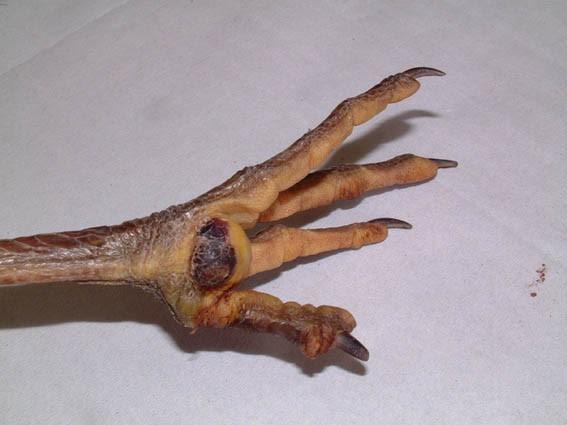
Figure 33. "Severe bumblefoot (Grade VI ) in a grey heron (Ardea cinerea). Severe tendon damage of the first digit was also present, and euthanasia was warranted in this case” (image credit Glen Cousquer. Wound assessment in the avian wildlife casualty. Used with permission).
A bird’s inactivity in an enclosure, limiting its ability to fly, is a contributing factor. In a study by P.T. Redig (Ford: Raptor Medicine Master Class), raptors that were housed outdoors and were able to exercise did not develop bumblefoot, regardless of their perching surfaces. The group that was maintained indoors on the same diet developed bumblefoot irrespective of the perching material.5
For large birds and raptors housed in wire enclosures, the walls of the enclosure should be designed with horizontal bars or solid barriers to minimize the tendency for hanging from the wire. The selection of proper perch size, shape, and covering for a particular species of bird is very important. Perches wrapped with hemp rope or covered with Astroturf work well for most raptors. Falcons do best on flat shelves or block perches covered with short Astroturf or cocoa mats. Strict sanitation of the facilities and feet is important to minimize bacterial infections. Liquid bandage products work well for minor skin cracks or torn talon sheaths in raptors.5 The feet should be massaged with a healing product, such as coconut oil or Aloe Vera gel (J. Miesle).
Figure 34. Red-tailed hawk at “Wild Care” in Eastham, MA. One talon had to be removed due to a serious infection in the feet. He is on ball bandages. The caregivers are optimistic about his ability to be returned to the wild” (image courtesy Cape Cod Capecast).
In most cases, treatment involves the surgical removal of scabs and adjacent necrotic and purulent (pus-filled) tissue, followed by suturing to achieve healing by first intention (the wound is held together by a blood clot or sutures). Sometimes antibiotic-impregnated beads are placed within the wound cavity to improve the rate of healing. If the opening is larger, sutures are used along with hydrocolloid dressings to promote healing.18

Figure 35. Grade VII “Bumblefoot infection has spread across both of this bird’s feet. The areas affected look blackened” (image courtesy The Veterinary Expert; used with permission).

Figure 36. Grade VI “A discrete bumblefoot lesion, showing typical positioning at the weight-bearing position on the base of the foot.” (Discrete refers to a single lesion that is localized as opposed to diffuse in which there are multiple lesions present) (image courtesy The Veterinary Expert; used with permission).

Figure 37. “Chickens cope incredibly well with legs bandaged! This bird has had surgery for bumblefoot. The bandages cover the surgical site as well as allowing pressure relief to the feet whilst the area in question heals” (image courtesy The Veterinary Expert; used with permission).
“Captive waterfowl are also at increased risk for developing this condition because of their heavy-bodied nature and the amount of time they spend standing around rough, hard surfaces around pools or pens. Waterfowl may suffer penetrating wounds and bruises on the feet which lead to pododermatitis.”15
Figure 38. A silver gull with a tibiotarsus fracture on its right leg (Larus michahellis) His solid left foot, burdened with more weight than normal, resulting in pododermatitis (image courtesy Kübra Gerbaga Özsemir).
Pododermatitis is common in poultry. VAPs (Vascular Access Ports) are more commonly used in mammalian patients but are sometimes used to treat avian patients requiring long-term intravenous therapy or serial blood collection. Vascular Access Ports offer the advantages of ease of access, reduced trauma and handling of the patient, and the accurate delivery of large volumes of tissue-irritating drugs. Although this technique is used for mammals, it is still considered a novel treatment for avian species.7

Figure 39. Grade VI bumblefoot foot in a buzzard (Buteo buteo). This bird had been shot and had developed severe arthritis of the tibiotarsal joint of the contralateral limb. The bird was emaciated and had a heavy worm burden. Excessive weight bearing on the healthy leg, coupled with malnutrition, is likely to have resulted in the lesions depicted (image courtesy World Wide Wounds).

Figure 40. Grade VI pododermatitis in a chicken (image courtesy Monica Talbett; used with permission).

Figure 41. Grade VI pododermatitis. The chicken’s foot before surgery (image courtesy of The Chicken Chick).

Figure 42. The chicken’s foot four months after surgery (image courtesy The Chicken Chick).
Case Studies involving Raptors
Case Study I: Technique: Advancement of a Skin Flap as a Novel Treatment for Pododermatitis in a Red-tailed Hawk, Sander, et.al., JAMS (27), 2013
A novel technique for healing of Stage VII pododermatitis in a captive Red-tailed hawk was discussed in the Journal of Avian Medicine and Surgery by Samantha Sander, et.al. of Urbana, Ill. In this case, a captive hawk presented with an inability to fly, and upon examination it was noted that a severe case of pododermatitis was present. This was due in part to the previous amputation of the second digit on the right foot, forcing the bird to put enough stress on the left foot to cause a tibiotarsal fracture. Over the next several months, the infection progressed from Grade I to Grade VII, and a multitude of treatments and preventive measures were taken, including medical and surgical treatments and environmental strategies. The left foot healed significantly, but the damage was done to the right foot during that time. The lesions on the right foot reached Grade VII, and despite treatment, were consistently more severe than the lesions on the left foot.19
As the condition of the right foot deteriorated, additional treatments were utilized, including surgical debridement and closure of wounds. Aerobic culture yielded Streptococcus species, Pseudomonas species, and Coliform bacteria. During this time, the hawk stood primarily on his left foot. Seven and a half months after admission to the clinic, the primary pododermatitis lesion on the right foot encompassed the entire metatarsal pad. Ulcers developed on the skin and soft tissues on the plantar surface of the foot, and the tendon sheath was exposed.19
Longer, more complex surgery followed. The wound was debrided and all diseased tissue was removed. The granulated tissue was resected (surgically removed), and a single flap was created using the skin tissue between the remaining digits. The edge of the flap was advanced over the affected tissue, covering the entire lesion with healthy foot skin. The area was padded and bandaged, and a bandaging cast was applied to reduce pressure on the flap during weight bearing.19
At first, it seemed that the wound was healing well; there was scabbing with partial dehiscence (opening of the wound), but that was managed; however, after four weeks, the area still had not healed. More debridement and treatment followed, and there was some improvement. Since the wound was healing appropriately, it was released to a wildlife rehabilitator for conditioning and evaluation of release status. But because the bird was forced to perch at a slight angle because of the missing digit on the other foot, it could not land on perches or grasp items. It was then euthanized because of its inability to apprehend live prey. This is the first time this new skin-flap technique was used.19

Figure 43. “Grade V pododermatitis lesion encompassing the metatarsal pad of the right foot of a red-tailed hawk before surgery. The hawk experienced a traumatic amputation of digit 2 of the right foot before admission. The digit was amputated later at the metatarsal-phalangeal joint” (images courtesy Sander: Advancement of a novel skin flap as a treatment for pododermatitis in a red-tailed hawk, AAV: JAMS, [27] 2013).”
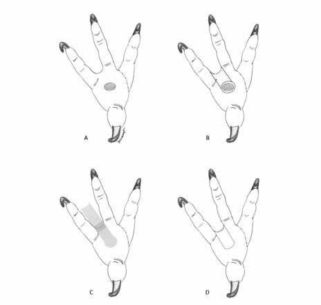
Figure 44. “Pictorial re-creation of the advancement flap procedure in a red-tailed hawk. (A) The lesion as it appeared before surgery. The ulcerate pododermatitis lesion is represented by the shaded circle at the level of the metatarsal pad, and the amputation site of digit 2 is represented by a hashed line. (B) Bilateral incisions (dashed lines) from the axial and abaxial edges of the debrided wound bed are made to develop a pedicle advancement flap by using the interdigital web skin between the third and fourth digits. The incisions extend over the web dorsally (not shown). (C) The pedicle advancement flap is created by elevating the skin from the subcutaneous tissue (light shading) and splitting the interdigital skin (dark shading). (D) The pedicle advancement flap is then used to cover the defect and is secured in place over the recipient bed.”
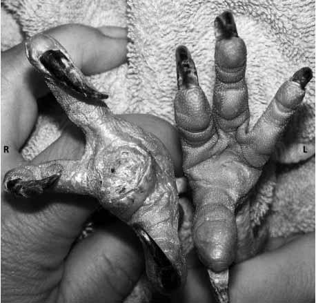
Figure 45. “The appearance of the surgical site 6 weeks after a pedicle advance flap was sued to surgically manage an advanced chronic pododermatitis lesion in a red-tailed hawk.”
Case Study II: Technique: Use of a Vascular Access Port for Antibiotic Administration in the Treatment of Pododermatitis in a Chicken, Doneley, AAV: JAMS (29) 2013
In this case, a chicken was presented to Dr. Robert Doneley of Queensland, Australia with chronic pododermatitis in one foot. It had been treated conservatively at first, but later surgically. Various antibiotics had been used, and several attempts at bandaging with antibiotic-impregnated beads had been made, both with poor outcomes. Cultures revealed E coli infections and Klebsiella species. The E. coli infection was resistant to most antibiotics. The plantar surface contained deep ulcerations and swelling, and the bird was moderately lame. The foot was radiographed and swabbed for sampling. The radiographs showed moderate degenerative joint disease in the bones of the foot. The bird was sent home with instructions to clean the wounds daily and to give the bird antibiotics and painkillers on a daily basis.7
Because the medications were not working as well as expected, the decision was made to implant a Vascular Access Port (VAP) in the right jugular vein to facilitate intravenous administration of the drugs. The chicken was hospitalized for two days and then was sent home with the owner with instructions on how to deliver medications through the port, cleanse the wounds, and change the bandages. Cultures were performed every two weeks, and after three consecutive negative culture results, treatment was discontinued.7
After three months, the wound was healed; however, severe degenerative arthritis was present in many of the foot joints. This was managed with medications. Since there was no evidence of infection, the VAP was removed, and the bird went on to live a normal life with its medications.7
Trauma to the plantar aspect of the foot leads to secondary infections with pathogens. These include Staphylococcus. aureus, Staphylococcus. epidermidis, Corynebacterium species, E. coli, Streptococcus faecalis, Pseudomonas species, Bacteroides species, Clostridium species, Candida albicans, and Aspergillus species.7

Figure 46. “Placement of a vascular access port in a domestic chicken for administration of long-term intravenous antibiotics. The right jugular vein (arrow) has been identified and mobilized.”

Figure 47. “Same as Figure 1. A tunnel has been created (arrows), connecting the incisions made for the placement of the port chamber and the catheterization of the jugular vein.”

Figure 48. Same as Figure 1. The “peel-away” sheath is removed after the intravenous cannula (arrow) has been introduced into the jugular vein.”

Figure 49. Same as Figure 1. Suturing the port chamber to the subcutaneous fascia.”

Figure 50. “Same as Figure 1. The vascular access port in situ after skin incisions have healed”
Appendix B: Food choices for affected birds
Vitamins should always be given to birds unless they are on a pelleted diet. These birds should be taken off pellets and put on a more natural diet. Avi-era is a good choice for vitamins. It can be purchased online or at some bird shops.
These foods are appropriate for other small birds like budgies, lovebirds, and medium and large birds as well. I buy everything separately and make my own mix. Don’t buy from big box stores or dog/cat pet stores. These stores may have had the food sitting in a warehouse for months or even years. Even if they sell birds, they buy from the big box stores. With bird-only stores, they buy quality, and you don’t run the risk of having contaminants like mold, animal droppings, and saliva in the food.
Don’t hesitate to eat with your bird. Many birds will start eating good foods if fed at the table with the owner and/or other birds. It’s a great bonding time.
Cereals:
- Sugar-free cereals, like Wheat, Rice, and Corn Chex; some like it moistened with a little rice, almond, or other non-dairy milk.
- Flakes, like Wheaties or cornflakes; Cheerios, LIFE cereal. Some may have small amounts of sugar, which is OK, but if sugar is among the first 3 ingredients, the food will have a lot of sugar in it.
- Flax seed—available at grocery stores, only for small birds
- Low-sugar granola. E.g., Quaker Puffed Granola, Blueberry-Vanilla Flavor. It has small freeze-dried blueberries in it.
Other foods:
- Nutriberries and Avi-cakes; are labeled as treats, but they are not. They are nutritious foods. They come in three sizes.
- Kaytee parakeet Forti-diet Honey Treat Bars for small to medium size birds. Break them up and give a small amount per day. Don’t just put the whole thing in the cage. Some of these birds will eat nothing else if allowed. Parakeet bars do not have sunflower or safflower in them, but the larger sizes do. https://www.chewy.com/kaytee-forti-diet-pro- health-honey/dp/122876.
Available at Chewy, Amazon, and Petco, among others.
Seeds
- Parakeet seed instead of cockatiel. They are the same, except that cockatiel includes a lot of sunflower and safflower, both bad for the liver. Read the ingredients on everything you buy.
- Oat groats: Some grocery stores carry it. Great Companions and Shiloh Farms do. I’ve been getting it at Shiloh. Good quality and price. Whole Foods or other similar stores may carry it. Amazon carries some good-quality groats. Do not buy anything cheap.
- Red and white/yellow proso millet:
White/yellow is available at some grocery stores or over the internet or at Morning Bird. Red and white both from Morning Bird. https://morningbirdproducts.com/collections/bird- seed, https://www.morningbirdproducts.com/products/morning-bird-red-proso- millet?_pos=1&_sid=937075e06&_ss=r
https://www.shilohfarmsmarket.com/ https://www.shilohfarms.com/
Never buy seeds from non-avian pet shops or big-box stores. Their seeds are stored for months or longer and may be contaminated with animal droppings or other dirt.
Millet sprays:
- Amazon, Nemeth Farms, has two bluebirds on top of millet. From “Bird Dog” Excellent quality. Available in 1 lb. bags and 5 lb. boxes. It’s quite expensive, but you get what you pay for. There is very little waste, and the sprays are full and fresh. It looks like this:
https://www.amazon.com/Nemeth-Farms-Parakeet-Natural- Healthy/dp/B001LK91O4/ref=sr_1_2?keywords=spray+millet+for+birds&qid=1567312878&s=ga teway&sr=8-2
https://www.amazon.com/Nemeth-Farms-Parakeet-Natural- Healthy/dp/B0068RFAFO/ref=sr_1_1?dchild=1&keywords=nemeth+farm+millet&qid=16088633 59&s=pet-supplies&sr=1-1
These websites have changed to offering the millet in 1-pound bags, not the 5 and 25 lb. boxes I wanted to get. So I went to another distributor in Amazon and found the 5 and 25 lb. boxes. Here is the link.https://www.amazon.com/Pesticide-Nemeth-Supplement-Cockatiels-Finches- 5lbs/dp/B001LK91O4/ref=sxts_rp_s_sp_1_0?content-id=amzn1.sym.497ddbaf-dbdc-4a61-b204- 33f5c59012b2%3Aamzn1.sym.497ddbaf-dbdc-4a61-b204- 33f5c59012b2&crid=2UGGL33HCZTNM&cv_ct_cx=millet%2Bspray%2Bfor%2Bbirds&keywords=millet% 2Bspray%2Bfor%2Bbirds&pd_rd_i=B001LK91O4&pd_rd_r=143c599f-3eb0-442a-a40b- 8c6cb500d950&pd_rd_w=xCxPs&pd_rd_wg=9ywgo&pf_rd_p=497ddbaf-dbdc-4a61-b204- 33f5c59012b2&pf_rd_r=D8V565J9WBVWKQZSVSQK&qid=1669429901&sprefix=millet%2Bspray%2B% 2Caps%2C73&sr=1-1-5985efba-8948-4f09-9122-d605505c9d1e&th=1
- Buckwheat seeds and groats, Meijers sometimes carries it, best price at Shiloh farms.https://www.shilohfarms.com/buckwheat-groats-organic/
- Zupreem fruit-flavored pellets for cockatiels and medium-sized birds, Chewy carries them; a few a day if they like them. It’s not for a nutritional benefit; simply if they like them.
- Kaytee Fruit and Veggie mix and Fruit Mix for Parakeets, and other parakeet mixes. Chewy, Petco, or Amazon.
- NO GRIT, ever, for psittacines birds (birds in the parrot family). These birds shell their seeds; only birds that eat their seeds whole use grit. Those birds, like canaries, doves, and pigeons, only need a very small amount; more should only be added when that amount is gone. Otherwise, they can get crop impaction.
Human foods:
- Rice, pasta, mashed potatoes, sweet potatoes, couscous, scrambled egg, applesauce (a real favorite, and a way to get fruit into them.) It’s also a good way to train them to eat from an oral syringe. Human foods are OK as long as they are not fatty, fried, or breaded. Baked chicken breast, ground beef, roast beef; deli, low sodium ham; and fish are also good. Usually, only the larger birds eat meat, but some smaller ones will try it.
- Tomato sauce (plain or mildly spicy), for spaghetti or on ground beef, small amounts
- Peas, corn, and other vegetables: fresh, green beans, asparagus, cauliflower, and broccoli, all lightly steamed or cooked. Cauliflower and broccoli can be given raw but there’s more nutritional value in them steamed. Even though the “raw diet” is popular right now, it is not always good for them. Most vegetables need to be cooked to release their nutritional value, and some birds have digestive issues when fed raw vegetables.
No raw sweet potatoes. They are toxic to birds. Serve only the fleshy meat inside; no seeds, stringy parts by the peel, or the peel itself. This information comes from a food chemist. No yams at all, ever. Don’t confuse yams with sweet potatoes. (T. Miesle, Food Chemist).
- Whole Foods, and similar stores, have different seeds and grains. Some they like; some they don’t. Try different foods with them.
- Whole grain bread. Oat-nut is a favorite. Quality white bread is good-many like their bread toasted.
- Soft cheese, like American cheese, occasionally. No other types since some have mold on them (like blue cheese). No dairy products, or rarely. I know many people like to give yogurt, but yogurt is dairy; if you give it, give small amounts and not often. They can’t digest lactobacillus.
Organic: "Organic" is not really different from regular food. Every state has different rules and regulations about what farmers can and can't use on their crops. And many of the foods we get are imported, where there are no regulations. Finally, once the food leaves the farm, there is no control at all as to what happens to it. It is handled by many middlemen between the farm and your table. (T. Miesle, Food Chemist).
Appendix C: The use of lasers in the treatment of pododermatitis
In 2014, ten veterinarians met during an Association of Avian Veterinarians conference to discuss the use of therapeutic lasers in treating avian and other exotics patients. Lasers are being used in veterinary medicine with positive clinical results, and low-power therapeutic lasers have proven effective for the treatment of wounds, reduction of inflammation, and modulation of pain. 17 The results of the discussion are as follows:
- All of the attending clinicians had used therapeutic laser treatment with birds and other exotics in their clinical work.
- These uses included:
- Healing wounds that have resisted previous therapies
- Painful conditions
- Wounds and skin infections
- Postsurgical incision treatment
- Osteoarthritis
- Pododermatitis
- Any inflammatory condition or traumatic lesion.7
When asked about the kind of response they observed with therapeutic laser treatment compared to routine care, the practitioners responded positively:
- One practitioner responded that several cases of pododermatitis in small mammals and birds had improved, in some cases dramatically, with the addition of therapeutic laser treatments
- Another mentioned that he had seen faster healing, a decrease in the severity of his wound cases, and improved mobility in orthopedic cases.
- A third mentioned accelerated healing time, particularly in the dermal wound healing of the foot.
- Another clinician saw considerable improvement in a chicken with bumblefoot, both in improved weight-bearing and decreased inflammation after just one treatment.
- One veterinarian found that laser treatments greatly reduced the bacterial colony in a wound.
- Another clinician noted that the animals appear to tolerate the treatment very well and are even able to relax a little during the treatment.7

Figure 51. “A cockatiel with a chronic, long-term self-mutilation site responded to his one session two weeks ago and received another one just in case. We had performed a complete set of appropriate tests on him over the last two years including bloodwork and radiographs, as well as a removal and biopsy of the affected skin to rule out damaged nerves” (image courtesy Vanessa Rolfe, The Bird and Exotic Hospital; used with permission).
A discussion of the advantages and disadvantages of therapeutic laser treatment in avian patients yielded the following results:
- The benefits:
- An increase in the level of healing for pododermatitis patients
- An adjunct to medical therapy alone
- Improved speed of treatment and response time, leading to less stress on the patient and better recovery and survivability of the patient.17
- The drawbacks:
- Difficulty in objectively determining the effectiveness or success of the laser treatments
- The lack of specific protocols for the use of the laser
- The large amount of mis- or disinformation being spread, which serves to confuse the clinician
- The lack of scientific studies in the literature about laser use. Those that are published sometimes have limited access, so clinicians may not have been able to read them and thus may not feel comfortable with their use.17
All concluded that the advantages significantly outweighed the disadvantages. Although lasers have been used for some time by veterinarians, they have not been used as long by avian and exotic veterinarians. All agreed that the differences between avian tissue and mammalian tissue warranted a more careful study of the techniques involved and results gleaned from such use, and far more care needs to be taken when applying laser treatment to birds.17
About
How to reference this publication (Harvard system)?
(2020) “Pododermatitis (Bumblefoot): Diagnosis, Treatment, Resolution and Prevention”, Avian Health and Disease. Available at: https://www.ivis.org/library/avian-health-and-disease/pododermatitis-bumblefoot-diagnosis-treatment-resolution-and-prevention (Accessed: 05 June 2024).

Comments (1)
Ask the author
1 commentThank you for a wonderfully interesting and well-illustrated review!