
Peridental Anatomy: Sinuses and Mastication Muscles
Author(s):
V.S. Cox
In: AAEP Focus Meeting - Focus on Dentistry - Albuquerque, 2011 by American Association of Equine Practitioners
Updated:
SEP 20, 2011
Languages:
Read
Take Home Message
The paranasal sinuses are closely related to the caudal maxillary cheek teeth and several cranial nerve branches. Dental disease can spread to these sinuses leading to nasal discharge and the possibility of serious neurological complications. Access to the sinuses should avoid damage to the infraorbital canal and nasolacrimal duct.
Introduction
Peridental entities such as the paranasal sinuses and mastication muscles play important roles in dental disease processes. This presentation is intended to describe the anatomy of these subjects.
The paranasal sinuses of the horse are formed by extension of the lining of the nasal cavity between external and internal plates of several skull bones.1 Much of this process occurs after birth.1 The frontal and maxillary bones1-7 are the main ones “excavated”1 to form the paranasal sinuses, but also, the sphenoidal and palatine bones are involved in sinus formation.6
Additionally these sinuses invade the dorsal, ventral and ethmoidal conchae (turbinates). Like the nasal cavity, right and left sinuses don’t communicate.1 The maxillary sinus of the horse is large and divided into rostral and caudal compartments.1-5,7 The facial crest is a landmark for the maxillary sinuses which lie between the boney orbit and the infraorbital foramen and deep to the facial crest.
The paranasal sinuses drain into the middle nasal meatus via slit like nasal maxillary apertures8-10 that come from both the rostral and caudal maxillary sinuses. The frontal sinus of other species communicates directly with the nasal cavity, but the equine conchofrontal sinus communicates with the nasal cavity indirectly by way of the caudal maxillary sinus.1 While the nasomaxillary apertures are thin and cryptic, the frontal maxillary opening is a large oval passage that is easily seen. The conchofrontal sinus drains into the dorsal aspect of the caudal maxillary sinus and the small sphenopalatine sinus drains into the ventral part of the caudal maxillary sinus.7
In the horse the main mastication muscles are the masseter and pterygoid muscles while the temporal and digastric muscles are of lesser significance.11,12 The masseter muscle lies lateral to the mandible while the medial and lateral pterygoid muscles attach to the medial side of the mandible. The medial pterygoid muscle is considerably larger than the lateral pterygoid muscle and the mandibular nerve passes between them.12 The latter muscle attaches close to the temporomandibular joint. Grinding movement of the cheek teeth is the result of synergistic action of the masseter and pterygoid muscles acting together to cause lateral/medial movement of the mandible.12 The masseter muscle has superficial and deep parts. The superficial part attaches to the ventral and caudal margins of the mandible while the deeper part has more vertical fibers and attaches to the lateral side of the mandible.12
Materials and Methods
In addition to traditional dissection methods, transverse bandsaw sections of cadaver heads were made to correlate with computed tomographic (CT) images from a live horse. Lateral radiographs of hemisected cadaver heads were used to create images free of overlapping structures on right and left sides.
Results
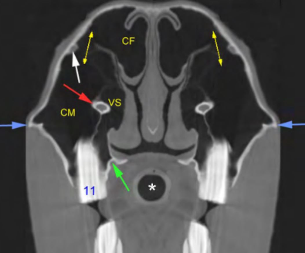
The frontal sinus of the horse is continuous with the dorsal conchal sinus forming a conchofrontal sinus. This sinus is the most dorsal of the paranasal sinuses; the maxillary sinuses are more ventral and lateral. The conchofrontal sinus communicates with the caudal maxillary sinus via the large oval frontal maxillary opening (Fig. 1) that lies medial and slightly rostral to the boney orbit.
Figure 1. CT section of the head of a live 7 year old Warmblood horse at the level of the last cheek tooth (11). CF = conchofrontal sinus, CM = caudal maxillary sinus, VS = vental conchal sinus, asterisk = lumen of endotracheal tube, arrows as follows, yellow = in the frontomaxillary opening, white = nasolacrimal duct, blue = facial crest, red = infraorbital canal, green = groove for great palatine artery which supplies the hard palate.
The dorsal concha was clearly attached to the other ethmoidal conchae (Fig. 2). The middle concha contained a space that opened to the caudal maxillary sinus or was not open. The ventral concha was not related to the ethmoidal conchae (Fig. 2).
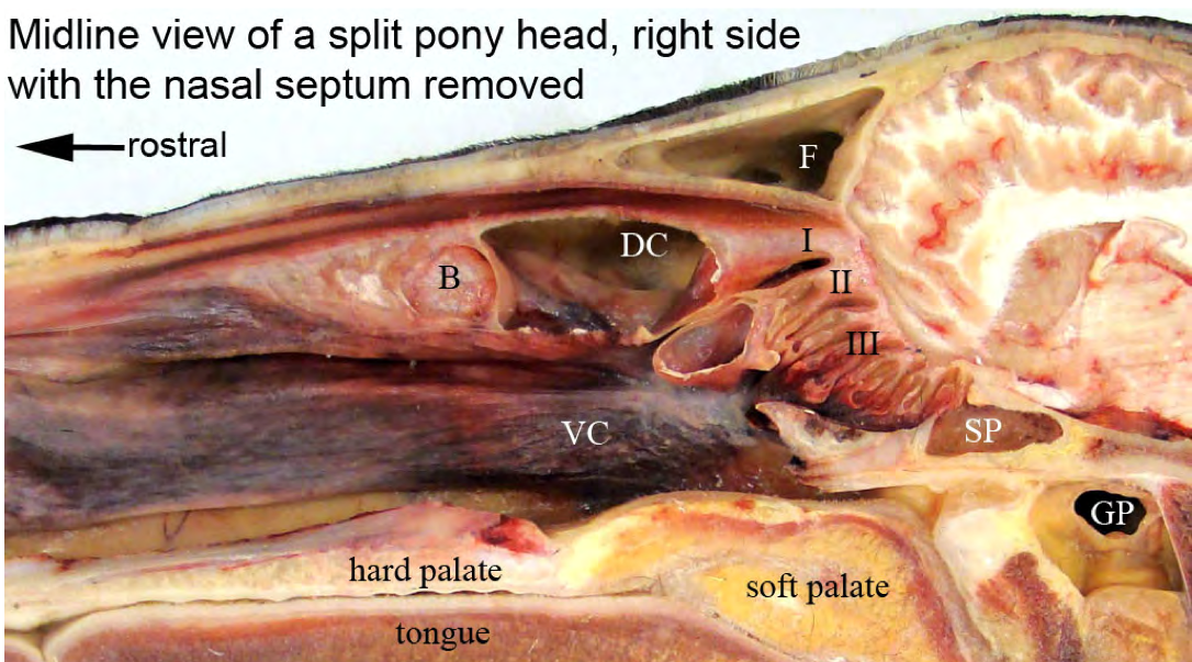
Figure 2. Split head of pony with windows cut into the dorsal concha and ethmoconcha II. I = ethmoconcha I (dorsal concha), II = ethmoconcha II (middle concha), III = ethmoconcha III, B = bulla in dorsal concha, DC = dorsal conchal sinus, F = frontal sinus, GP = guttural pouch, SP = sphenopalatine sinus, VC = ventral concha.
The caudal maxillary sinus is considerably larger than the rostral maxillary sinus. The size of both maxillary sinuses is increased significantly with age as the maxillary cheek teeth shorten due to attrition. In the young animal the infraorbital canal is in direct contact with the apical part of the alveoli of the caudal cheek teeth (Fig. 3) but with age the distance between the infraorbital canal and the alveoli increases and a thin plate of bone connects the canal to the dental alveoli (Fig. 1). The alveoli of the caudal cheek teeth and the infraorbital canal form a wall that separates the rostral maxillary sinus from the more medial ventral conchal sinus (Fig. 3).
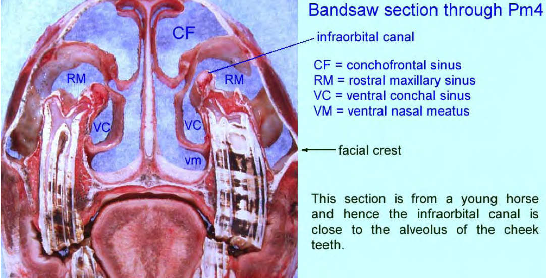
Figure 3. Bandsaw section through the rostral maxillary sinus.
[...]
About
Copyright Statement
© All text and images in this publication are copyright protected and cannot be reproduced or copied in any way.Provided by:

Comments (0)
Ask the author
0 comments