
Ultrasonographic Imaging of the Adult Equine Acute Abdomen
Author(s):
In: AAEP Focus Meeting - Focus on Colic - Indianapolis, 2011 by American Association of Equine Practitioners
Updated:
JUL 26, 2011
Languages:
Read
Take Home Message
This paper will describe a rapid, systematic examination protocol for ultrasound evaluation of the adult colic patient. Once the clinician is comfortable with the normal ultrasonographic anatomy and alterations commonly seen in the wide spectrum of colic disorders, they should be able to quickly categorize the condition into gastric, small, or large intestinal disorders in order to add important information to the total colic examination procedure. The overall objective is to provide the horse owner with a more rapid and accurate diagnosis and recommended treatment for their horse.
Introduction
Besides the physical examination, ultrasound is the author’s next step in the evaluation of the critical colic patient. Rectal examination remains useful, but is limited to structures that are within of the examiner in the caudal abdomen. The abdominal cavity reaches cranially to the diaphragm just caudal to the heart. A systematic approach to the ultrasonographic evaluation of the abdomen allows for rapid assessment of location, characterization, and content of multiple intra-abdominal structures.
Understanding Ultrasound Equipment
A variety of options are available in the offerings of ultrasound equipment. Newer, more portable devices are coming on the market each year – both digital and in laptop configurations. Some understanding of the physics of ultrasound is necessary – mainly in understanding the principles of frequency. The higher frequency wavelengths provide more detailed images, but less (more shallow) tissue penetration. An example being a 12- mHz probe is usually used for tendon imaging… providing excellent image quality with fine detail, but unable to visualize anatomic structures more than a few centimeters deep to the skin. Alternatively, a 1.9-mHz probe provides adequate depth of penetration to image the entire equine heart, but does not produce the fine detail image of the high frequency probe. The type of probe used should be appropriate to the body region being evaluated. The smaller, microconvex probes are good for accessing small body parts, especially in tight confined areas such as the inguinal region. The curvilinear 3.5 mHz probe is probably the best all-around probe for general abdominal imaging of the colic patient. Various other probes may be used for additional evaluation (higher frequency, higher detail probes for more detail of the serosal surfaces, or lower frequency probes to penetrate large volume effusions or to evaluate the pregnant uterus).
Patient Preparation
Preparation of the area to be imaged is also an issue. Some coupling material is required for the sound waves to penetrate the hair and skin. For most situations, isopropyl alcohol alone is used to wet down the hair coat without clipping. This may degrade the surface material of the probe over time. On some occasions, clipping the hair and application of a contact gel will be required to achieve the best detail in the image.
The Ultrasound Exam
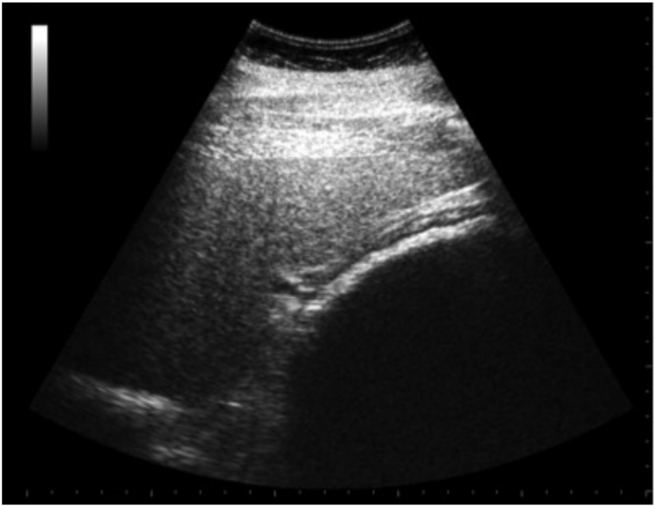
The practical part of the ultrasound examination is that it can be quickly performed immediately following the physical examination. In some situations, it can be performed prior to the rectal examination. For the abdomen, a systematic approach is helpful as with the physical examination. The author prefers a system starting cranially on one side – just caudal to the elbow, progressing caudally, imaging all appropriate structures on that side, and then repeating the process on the contralateral side. This way, a complete examination of the body cavity is performed in an effort to determine the nature of the disease process. On the left side, one would start near the elbow – scanning dorsal-to-ventral in each intercostal space to include the ventral lung margin and diaphragm. Most cranially - observing the liver, moving caudally to observe the stomach and its contents (mainly looking for excessive fluid volume as would occur with intestinal obstruction), and the spleen. The spleen-stomach relationship is important to identify on the left side (Fig. 1). Evaluation of the stomach content and size can be determined. The typical adult gastric profile is visible over 3-4 intercostal spaces. Colonic displacements or epiploic foramen entrapments can obscure the normal stomach-spleen image. Small intestinal segments are usually observed in the caudal-ventral abdomen just medial to the ventral margin of the spleen. Normal small intestine rarely contains luminal fluid, and will often be seen as variably motile structures between the medial surface of the spleen and the colon.
Figure 1. Spleen-stomach relationship on the left side.
[...]
About
How to reference this publication (Harvard system)?
(2011) “Ultrasonographic Imaging of the Adult Equine Acute Abdomen”, AAEP Focus Meeting - Focus on Colic - Indianapolis, 2011. Available at: https://www.ivis.org/library/aaep/aaep-focus-meeting-focus-on-colic-indianapolis-2011/ultrasonographic-imaging-of-adult-equine-acute-abdomen (Accessed: 24 April 2024).
Author(s)

Copyright Statement
© All text and images in this publication are copyright protected and cannot be reproduced or copied in any way.Provided by:

Comments (0)
Ask the author
0 comments